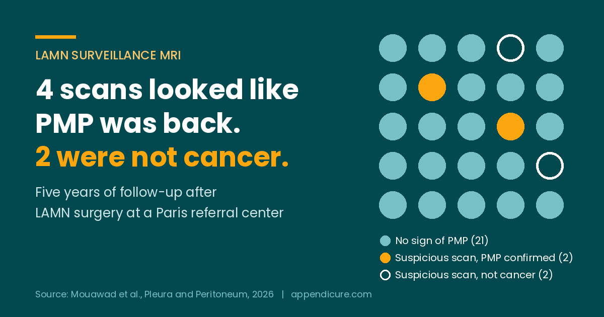

Short answer: LAMN surveillance MRI flagged both cases of pseudomyxoma peritonei (PMP) that developed during follow-up in a new five-year study from a Paris referral center. It also raised two false alarms. Of the four patients whose scans looked suspicious for PMP, two actually had it and two did not, and all four went back for major surgery. Blood tumor markers stayed normal the whole time, including in the two patients who really did develop PMP.
This study is about low-grade appendiceal mucinous neoplasm, or LAMN, found after an appendectomy, in people whose first scans and blood work after surgery were clean. It does not apply to HAMN, to people who already had PMP when they were diagnosed, or to any type of appendiceal adenocarcinoma, including goblet cell adenocarcinoma and signet ring cell.
Back in April I wrote about a LAMN patient whose follow-up plan felt like a placeholder. One of the studies I cited then was a small active surveillance program at the Pitié-Salpêtrière hospital in Paris, led by Professor Marc Pocard, with about three years of follow-up. That same team has now published five years of results in the journal Pleura and Peritoneum. The new paper is open access, so anyone can read it.
How the LAMN surveillance MRI program worked
The team enrolled 30 people between 2014 and 2021 who had LAMN on their appendix pathology. To get in, a patient needed a normal MRI after surgery, normal CEA, CA 19-9 and CA-125, and no more than a small amount of mucin outside the appendix. A tumor board reviewed every case and agreed that no further surgery was needed.
Instead of more surgery, each patient got a physical exam, the three tumor markers, and an MRI of the abdomen and pelvis at six months, then once a year. The plan runs for 10 years and stops at age 75. Five people passed 75 during the study and were dropped, so the five-year report covers 25 patients. Their median follow-up was just under five years, and some had quite a bit less.
These were not all the lowest-risk patients. On the pathology, 16 of the 25 had a perforated appendix. Two had cells in the mucin outside the appendix, and one had mucin with no cells. In 12 patients, the surgeon saw mucin around the appendix during the first operation.
What the scans found
Twenty-one of the 25 patients showed no sign of PMP on any scan. Four had an MRI that looked like the disease might be coming back. The findings were things like small pockets of fluid in the pelvis or around the liver and spleen, and small nodules. A tumor board reviewed each one and sent all four for cytoreductive surgery.
Two of those four really had low-grade PMP. Both had a perforated appendix at their first surgery, and one had cells in the mucin outside the appendix. Both suspicious scans came early, at six months and at one year. Surgeons removed all visible disease in both. One had bleeding inside the abdomen nine days later and needed another operation. The other had the disease return about two and a half years after the first cytoreduction and had a second one. Both showed no sign of disease on their most recent MRI.
The other two did not have cancer. One had a benign fluid-filled sac called a peritoneal inclusion cyst. The other had no tumor at all in anything the surgeons removed. One of them developed a hernia at the incision that had to be repaired a year later.
Nobody who was clear at the three-year check went on to develop PMP by the five-year check.
Why a scan can look like PMP when it isn’t
After abdominal surgery, the body heals with scar tissue, small fluid collections and inflamed patches of lining. On MRI, some of that can look a lot like early mucin deposits. The authors say directly that telling normal healing or harmless fluid apart from early PMP is one of the main weak spots of follow-up by imaging.
Because of their two false alarms, the Paris team changed what they recommend. They now say a single MRI should not be enough to send someone to cytoreductive surgery. When a scan is unclear, they suggest a CT scan plus a repeat MRI three to four months later to see whether the spots stay or grow. If PMP is still suspected, they suggest a diagnostic laparoscopy first. That is a small keyhole operation where a surgeon looks inside and sends tissue to the pathologist during the procedure, before anyone commits to the full CRS with HIPEC. They also advise placing those small incisions along the midline of the belly, because PMP can grow into port sites if the disease turns out to be real.
Please read this before you act on anything above
This study is about how to confirm a suspicious scan. It does not say to ignore one, and it does not say to skip scans. Both real cases in this study were flagged by MRI and confirmed at surgery, early enough that surgeons could remove all visible disease.
If your scan shows something unclear, the question for your team is how they plan to confirm it before surgery, and whether a repeat scan or a look with laparoscopy makes sense in your case. That is a decision to make with a surgeon who treats a lot of appendix cancer.
If you already have PMP, HAMN, or any adenocarcinoma, this study was not built for you. If your report shows cells in mucin outside the appendix, more extensive mucin outside the appendix, or a positive or unclear margin, your situation may not match the lower-risk patients in this study. That deserves its own surveillance conversation with your team.
The tumor markers never moved
The team checked CEA, CA 19-9 and CA-125 throughout. None of them went up in any patient, at any point, including the two who developed PMP. In this group, the MRI flagged the disease and the blood work did not.
Many LAMN patients in the Appendicure group are told their markers are normal and that this is good news. Normal markers are good news, but in low-grade disease they are not proof that nothing is there. The authors also admit some patients had gaps in their marker records, so this is not a perfect test of the markers. I have written more about what tumor markers can and cannot tell you, and about why CA-125 matters for men too.
How long LAMN follow-up should last
In this study, both real cases showed up within the first year. It would be easy to read that as a reason to relax after year one. I would not. This was 25 people, and only two of them developed PMP, which is too few to say when it tends to happen.
A larger structured surveillance program in Ireland followed 83 people with LAMN using yearly CT scans. That was a broader group than the Paris cohort, and 15 of them already had mucin inside the abdomen when they were diagnosed. Overall, 6 percent progressed to PMP, the median time to progression was 23 months, and the highest risk was in the first three years. Two of the 60 people with no mucin outside the appendix progressed, compared with 3 of the 15 who had it. So the 6 percent figure is not the risk for someone who looks like the Paris patients, and in the Irish group some cases came well after the first year.
Guidelines also disagree on how often to scan. The Paris team recommends LAMN surveillance MRI at six months after surgery and then once a year for at least 10 years. The 2025 German S2k guideline on LAMN recommends surveillance every six months for five years.
The limits of this study
This was a small study at one expert center, and the patients were carefully chosen by a tumor board that sees a lot of peritoneal disease. There was no comparison group, so it cannot say whether surveillance does better or worse than extra surgery up front. The results were only described, not tested statistically. The five people who aged out at 75 were not followed further. Many pathology slides came from outside hospitals, which sometimes limited how completely the specimen could be reviewed.
One detail in the paper doesn’t match up. The text says all four patients with suspicious scans had CRS with HIPEC, but the paper’s own table marks one of the two benign cases as not getting HIPEC. Either way, all four had major abdominal surgery.
The authors say they used ChatGPT to help with language editing, and they state they reviewed and take responsibility for the content. They report no outside funding and no conflicts of interest.
Questions to bring to your team
If you have LAMN and are in surveillance, these are fair questions for your next visit. What is my written schedule for scans and tumor markers, and for how many years? Will my imaging cover the abdomen and the pelvis, and is MRI or CT better for me? If a scan shows something unclear, what is your plan to confirm it before surgery? Would you repeat the scan first, or consider a diagnostic laparoscopy? And if I do need surgery, will it be at a center that does a lot of CRS with HIPEC?
Frequently asked questions
What is LAMN surveillance MRI?
LAMN surveillance MRI is regular MRI scanning of the abdomen and pelvis after a low-grade appendiceal mucinous neoplasm is removed. It watches for mucin or tumor spreading in the abdomen, which is called pseudomyxoma peritonei or PMP.
Can an MRI show PMP when there is none?
Yes. In a 2026 Paris study of 25 LAMN patients, 4 had MRIs that looked like PMP. Only 2 had PMP. The other 2 had a benign cyst or no tumor at all. Healing tissue and harmless fluid after surgery can look like early disease.
Can normal tumor markers rule out PMP during LAMN surveillance?
No. In the same study, CEA, CA 19-9 and CA-125 stayed normal in every patient, including the two who developed PMP. MRI flagged the disease. Markers are useful, but they do not replace imaging for LAMN.
What should happen if a surveillance scan looks suspicious?
The Paris authors now suggest not deciding on one MRI. They recommend a CT scan and a repeat MRI in three to four months, and a diagnostic laparoscopy with tissue checked during the procedure before committing to CRS with HIPEC. Talk this through with an experienced appendix cancer surgeon.
Does this study apply to HAMN or appendiceal adenocarcinoma?
No. It only included people with LAMN whose scans and tumor markers were normal after surgery. It does not cover HAMN, PMP present at diagnosis, mucinous or non-mucinous adenocarcinoma, goblet cell adenocarcinoma, or signet ring cell.
Read more
When Surveillance Feels Like a Placeholder: A Closer Look at LAMN Follow-Up After Appendectomy
What Tumor Markers Tell Us Before and After CRS
LAMN patients are the ones most often missing from research
This study had 25 people. The Irish program had 83. That is how thin the LAMN evidence still is. The Appendicure Patient-Led Global Appendix Cancer Registry collects pathology, molecular results and treatment from patients directly, so you do not need your hospital to enroll you. If you have LAMN, your scan schedule, your markers and any suspicious findings help build the picture the next patient needs.
Already enrolled? Add new scans or reports, or correct your record here.
Appendicure is a 501(c)(3) nonprofit, EIN 41-5040966. If you want to help keep this work going, you can donate here.
Sources
Mouawad C, Bardier A, Lucidarme O, Doat S, Djelil D, Fawaz J, Pocard M. Active surveillance strategy for low-grade appendiceal mucinous neoplasm (LAMN): 5-year results. Pleura and Peritoneum, published online September 14, 2026. doi.org/10.1515/pp-2025-0043
Mouawad C, et al. Active surveillance for low-grade appendiceal mucinous neoplasm (LAMN). Pleura and Peritoneum 2024;9:31-7. doi.org/10.1515/pp-2023-0032
Hannan E, et al. Surveillance of low-grade appendiceal mucinous neoplasms for progression to pseudomyxoma peritonei: results from a structured surveillance programme. Colorectal Disease 2025;27:e17266. doi.org/10.1111/codi.17266
Köhler F, et al. German S2k-guideline on diagnostics, treatment and surveillance of low-grade appendiceal mucinous neoplasms (LAMN). European Journal of Cancer 2025;222:115430. doi.org/10.1016/j.ejca.2025.115430
This post is for education, not medical advice. Decisions about surveillance and surgery for LAMN belong with a clinician who treats a lot of appendix cancer.
Leave a Reply