A plain-language guide to the types of appendix cancer for patients and caregivers reading a pathology report, comparing notes online, or trying to understand which diagnosis they have. Different centers and older reports use different names for the same thing, and that confusion is part of why this page exists.
Reviewed by appendiceal cancer specialists. Based on the 2025 Peritoneal Surface Malignancies Consortium consensus guidelines and the 2019 WHO Classification of Digestive System Tumors.
Appendix Cancer Is Its Own Disease
Appendix cancer is biologically, molecularly, and clinically distinct from colon cancer. That distinction matters because for decades, appendix cancer was treated as a rare offshoot of colorectal cancer, and treatment decisions were extrapolated from colon cancer research. The published appendiceal cancer literature has now made it clear that this approach has limits.
The molecular profile of appendiceal tumors differs from colorectal tumors in important ways. Mucinous appendiceal tumors carry KRAS and GNAS mutations at high rates and APC mutations rarely, the opposite pattern from most colon cancers. Response to chemotherapy in appendix cancer is generally lower than in colon cancer. Some types of appendix cancer do not benefit from standard colon cancer regimens at all. The biology is different, and the treatment has to follow the biology.
The current evidence-based reference
The 2025 Peritoneal Surface Malignancies Consortium consensus guidelines (Godfrey et al., published in Cancer) represent the current evidence-based reference for appendiceal cancer management. These guidelines were developed by 138 appendiceal cancer specialists through a modified Delphi consensus process, with over 90% agreement on all pathway recommendations. They are appendiceal-specific, not extrapolated from colon cancer.
Understanding Tumor Grade and Differentiation
Before getting into specific diagnoses, there is one piece of pathology vocabulary that shows up across nearly every appendix cancer report: differentiation. It is one of the most important findings on a pathology report and one of the most confusing for patients.
Differentiation describes how much the cancer cells look like normal, healthy cells when the pathologist examines them under a microscope. The closer the cancer cells look to normal cells, the more “differentiated” the tumor is. The less they look like normal cells, the more “undifferentiated” the tumor is. Differentiation is what the pathologist translates into a grade, written as G1, G2, or G3 on most reports.
The three levels of differentiation, in plain language
- Well-differentiated (Grade 1, G1). Cancer cells look very similar to normal healthy cells. The tumor still resembles the tissue it came from. Generally the slowest-growing and least aggressive. LAMN is classified as G1.
- Moderately differentiated (Grade 2, G2). Cancer cells look noticeably different from normal cells but still keep some recognizable features. Intermediate behavior between well-differentiated and poorly differentiated. HAMN is classified as G2.
- Poorly differentiated (Grade 3, G3). Cancer cells look very different from normal cells. The tumor has lost most of the features of the tissue it came from. Generally the most aggressive. Signet ring cell mucinous adenocarcinoma (more than 50% signet ring cells) is classified as G3.
- Undifferentiated (sometimes called “GX” or grouped as G3). Cancer cells look so abnormal that the pathologist cannot identify what type of tissue they came from. This is the most aggressive category and is the defining feature of Undifferentiated Carcinoma, NOS (UC-NOS).
The phrase “highly differentiated” is sometimes used informally to mean well-differentiated. It is not a standard AJCC grade category. If your report uses “highly differentiated,” it almost certainly means G1 (well-differentiated). If there is any uncertainty, ask your pathologist or oncologist which AJCC grade applies.
Why grade matters more than type in some situations
For metastatic mucinous disease (Stage IV with peritoneal spread), the grade is what determines the stage grouping, not just the tumor type. M1b disease with G1 is classified as Stage IVA. M1b disease with G2 or G3 is classified as Stage IVB. This is the only stage where grade is used in the stage grouping itself, and it reflects how strongly grade predicts behavior in metastatic disease.
Grade also shows up in the staging tables below. For a quick visual reference of how grade maps to AJCC categories, see the Tumor Grade section under Staging.
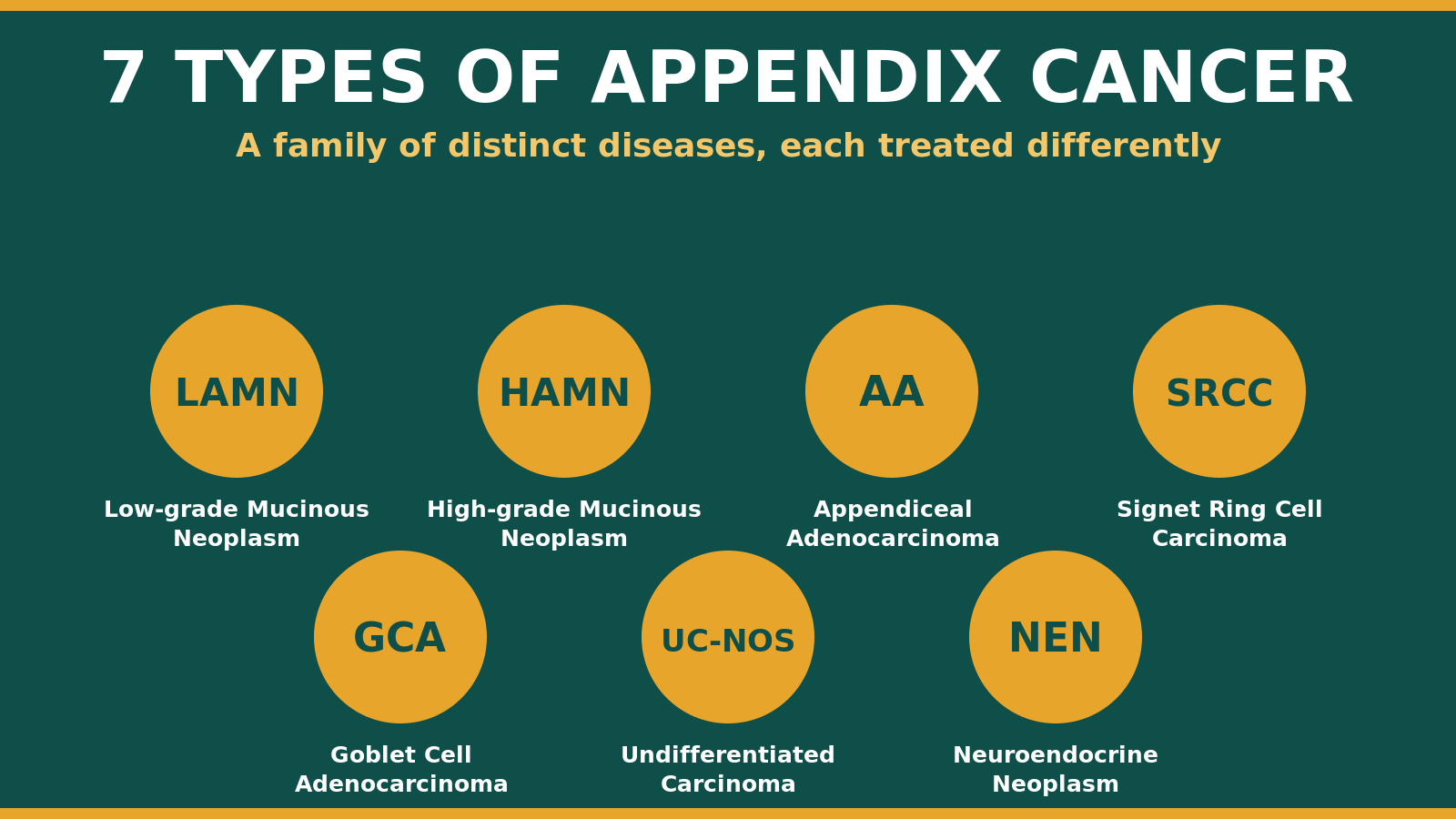
The Main Types of Appendix Cancer
Appendix tumors fall into several distinct categories. The most clinically important types of appendix cancer, and the ones this page covers in depth, are mucinous neoplasms (LAMN and HAMN), appendiceal adenocarcinoma in its several forms, goblet cell adenocarcinoma, and the rare undifferentiated carcinoma. Appendiceal neuroendocrine neoplasms are covered briefly because they are biologically separate and managed under different guidelines.
Use the jump menu above to skip directly to your type, or read through the section below to understand how the types relate to one another. For how each type is diagnosed and treated, see our diagnosis and treatment page, or start with our overview of what appendix cancer is.
Low-Grade Appendiceal Mucinous Neoplasm LAMN
Mucinous adenoma, mucinous cystadenoma, or mucinous neoplasm of uncertain malignant potential (UMP). If your pathology report uses any of these older terms, the diagnosis is the same condition now called LAMN.
What it is
A low-grade mucinous tumor of the appendix that grows by pushing through the appendix wall rather than invading destructively. LAMN is the most common origin of pseudomyxoma peritonei (PMP). It was named in 2003 to distinguish it from invasive cancer and from benign mucoceles.
How it behaves
Indolent when confined to the appendix. The main risk is perforation of the appendix wall, which spills mucus-producing cells into the abdomen and can lead to PMP. Even disseminated LAMN tends to grow slowly compared with adenocarcinoma.
Molecular features
KRAS and GNAS mutations are very common. Microsatellite stable. TP53 mutations are rare. This molecular pattern differs from colon cancer and is part of why LAMN responds differently to standard chemotherapy regimens.
Staging
LAMN has its own special staging code, pTis(LAMN), used when the tumor is confined to the appendix. If the tumor reaches the subserosa it is pT3. If acellular mucin is found outside the appendix it is pT4a. If cellular mucin or peritoneal disease is found it is staged as M1b. The distinction between acellular mucin (mucin without tumor cells) and cellular mucin (mucin containing tumor cells) is one of the most consequential findings on a pathology report. Acellular mucin has the best prognosis. Cellular mucin indicates that tumor cells have already escaped the appendix.
How it is treated
For LAMN confined to the appendix with negative margins, no perforation, and no extra-appendiceal mucin, appendectomy alone is the recommended treatment, followed by selective surveillance. This is supported by 96% consensus in the 2025 guidelines.
For LAMN with microscopic perforation or extra-appendiceal mucin confined to the appendix surface, appendectomy plus surveillance is recommended (98% consensus).
For LAMN with positive margins where viable tumor cells (not just mucin) remain at the resection edge, the recommendation is the most conservative additional surgery possible to achieve negative margins. The consensus specifically recommends against routine ileocecectomy or right hemicolectomy for LAMN. This is a meaningful shift away from older practice patterns that defaulted to hemicolectomy.
For LAMN with disseminated acellular mucin or any cellular mucin in the abdomen, the peritoneal disease pathway applies and cytoreductive surgery becomes part of the discussion.
↑ Back to topHigh-Grade Appendiceal Mucinous Neoplasm HAMN
HAMN is a newer category formally recognized in WHO 2019. Some older reports may have classified the same lesions as mucinous neoplasm of uncertain malignant potential (UMP) or grouped them with mucinous cystadenoma with high-grade features.
What it is
A mucinous tumor of the appendix with the same pushing-type growth pattern as LAMN, but with high-grade cytologic features. The cells look more abnormal under the microscope. HAMN is rarer than LAMN and less well characterized in the literature.
How it behaves
More likely to progress than LAMN, but does not show the destructive invasion of a true adenocarcinoma. Behavior in the peritoneal cavity, when it spreads, tends to follow patterns of other higher-grade mucinous tumors.
Molecular features
Similar to LAMN, but TP53 mutations are more common. The first dedicated clinicopathologic and molecular characterization of HAMN was published in 2026 (Seldomridge et al., Annals of Surgical Oncology).
How it is treated
The 2025 consensus treats HAMN with largely the same algorithm as LAMN. The key difference is for localized acellular extra-appendiceal mucin, where cytoreduction can be considered rather than observation alone, given higher rates of progression. The consensus also does not recommend routine right hemicolectomy for HAMN in the absence of peritoneal disease. This is a published divergence from some older international guidelines that did suggest hemicolectomy for HAMN.
↑ Back to topAppendiceal Adenocarcinoma AA
The mucinous form was historically called mucinous cystadenocarcinoma. You may also see references to colonic-type adenocarcinoma of the appendix for the non-mucinous subtype. These older terms describe what is now classified as appendiceal adenocarcinoma.
What it is
A true cancer of the appendix that invades destructively into surrounding tissues. The key pathologic distinction from LAMN and HAMN is the type of growth: AA shows destructive infiltration and desmoplasia (a fibrous tissue reaction to invading tumor), where LAMN and HAMN push but do not invade in this way.
Subtypes within AA
Appendiceal adenocarcinoma is not a single disease. It includes:
- Mucinous adenocarcinoma. The most common AA subtype. Produces large amounts of mucin and frequently spreads through the peritoneal cavity.
- Non-mucinous (intestinal-type) adenocarcinoma. Behaves more like colon cancer in growth pattern and metastatic spread to liver and lymph nodes.
- Mucinous adenocarcinoma with signet ring cell component (50% or fewer signet ring cells). An intermediate-aggressiveness subtype.
- Mucinous signet ring cell carcinoma (more than 50% signet ring cells). The most aggressive AA subtype. Covered separately below.
Non-Mucinous (Colonic-Type) Adenocarcinoma
This is the form of appendiceal adenocarcinoma that does not produce significant mucin. Under the microscope the cells and growth pattern resemble a typical colon cancer, which is why it is also called colonic-type or intestinal-type adenocarcinoma. Older pathology reports sometimes use the term colonic-type adenocarcinoma of the appendix.
It tends to behave more like colon cancer than the mucinous types do. It is more likely to spread through the lymph nodes and to the liver, rather than producing the mucin-driven peritoneal spread seen with mucinous tumors. Non-mucinous histology is treated as a high-risk feature when planning treatment. Care follows the appendiceal adenocarcinoma pathway above, and tumor grade remains central to decisions about chemotherapy.
Molecular profile
Common mutations include KRAS, GNAS, TP53, SMAD4, PIK3CA, and APC. APC mutation is notably less common than in colon cancer. Microsatellite instability is rare (around 1 to 6 percent of cases). Molecular profiling of AA has identified at least four distinct subtypes (Foote et al., 2023, Journal of Clinical Oncology): a RAS-mutation-predominant group with relatively favorable outcomes, a GNAS-mutated group associated with treatment resistance and high peritoneal disease burden, a TP53-predominant group with chromosomal instability and poorer outcomes, and a fourth subtype defined in the same paper. These molecular subtypes are currently most useful for research and clinical trial design.
How it is treated, localized disease
Right hemicolectomy with regional lymphadenectomy (at least 12 lymph nodes for accurate staging) is the standard surgical treatment for AA confined to the appendix. This is supported by 97% consensus.
An exception applies for well-differentiated (Grade 1) mucinous adenocarcinoma confined to the appendix with negative margins, where observation rather than hemicolectomy may be appropriate.
For Stage III disease, or Stage II with high-risk features, adjuvant systemic chemotherapy can be considered (98% consensus). High-risk features include T4 stage, poor differentiation, bowel obstruction or perforation, vascular, lymphatic, or perineural invasion, fewer than 12 lymph nodes examined, lymph node involvement, signet ring cell features, and non-mucinous histology.
For Stage I or II without high-risk features, surveillance is recommended without adjuvant chemotherapy.
An important nuance on adjuvant chemotherapy
Recent appendiceal-specific research has found that across AA subtypes, including goblet cell adenocarcinoma, adjuvant chemotherapy after surgery for early-stage disease has not shown a survival benefit. The full manuscript is in submission. This is a meaningful divergence from the colon cancer standard that has historically influenced AA management. Patients with early-stage disease should ask their treatment team about appendiceal-specific data rather than colon cancer extrapolations when discussing adjuvant chemotherapy.
How it is treated, peritoneal disease
For AA with peritoneal spread, the 2025 consensus recommends upfront systemic chemotherapy to assess disease response, followed by planned complete cytoreductive surgery with intraperitoneal chemotherapy if complete cytoreduction is achievable (95% consensus).
The consensus also notes that right hemicolectomy does not confer survival benefit for Grade 1 mucinous AA with peritoneal spread when complete cytoreduction is feasible without it. This is an important distinction patients can ask about, because older practice patterns sometimes default to hemicolectomy even when the published evidence does not support it for this specific subgroup.
↑ Back to topSignet Ring Cell Carcinoma SRCC
You may see this written as mucinous signet ring cell carcinoma, SRCC, or mucinous adenocarcinoma with signet ring cell features. The diagnosis depends on what percentage of the tumor cells show the signet ring pattern (more than 50% versus 50% or fewer).
What it is
A subtype of mucinous adenocarcinoma defined by the appearance of the tumor cells under the microscope. The cells contain large pools of mucin that push the nucleus to one edge, creating a “signet ring” appearance. When more than 50% of the cells show this pattern, the tumor is classified as mucinous signet ring cell carcinoma. With less than 50%, it is mucinous adenocarcinoma with signet ring cell component.
How it behaves
The most aggressive form of appendiceal adenocarcinoma. Strong association with peritoneal spread, lymph node involvement, and distant metastasis. Outcomes are worse than for other AA subtypes.
How it is treated
The 2025 consensus considers signet ring cell histology a high-risk feature that warrants consideration of more intensive treatment, including systemic chemotherapy in some localized cases where it would not otherwise be recommended.
One important caution from pathology
Degenerating cells in low-grade pseudomyxoma peritonei can sometimes look like signet ring cells under the microscope, even though they are not true signet ring cells. The 2025 consensus pathology section notes this can produce a misleading diagnosis. If a pathology report describes signet ring cells alongside an otherwise low-grade primary tumor, expert review at a high-volume appendiceal cancer pathology service is worth pursuing before treatment decisions are made based on that finding.
Goblet Cell Adenocarcinoma GCA
Goblet cell carcinoid, adenocarcinoma ex-goblet cell carcinoid, mixed carcinoid-adenocarcinoma, or crypt cell carcinoma. All of these older terms describe what is now classified as goblet cell adenocarcinoma. The name change in WHO 2019 is meaningful because GCA is not a neuroendocrine tumor and should not be managed under neuroendocrine guidelines.
What it is
A distinct tumor type that contains features of both glandular tumors (adenocarcinoma) and mucin-producing goblet cells. In the past it was called “goblet cell carcinoid,” which implied it was a neuroendocrine tumor. WHO 2019 reclassified it as goblet cell adenocarcinoma to reflect that it is not a neuroendocrine tumor and should not be managed under neuroendocrine tumor guidelines.
Why the name change matters
If a pathology report uses the term “goblet cell carcinoid,” that is the older name for goblet cell adenocarcinoma. The diagnosis is the same condition, but the treatment approach is meaningfully different. Neuroendocrine tumor staging systems and neuroendocrine-targeted therapies (such as somatostatin analogs) should not be applied to GCA. Treatment follows adenocarcinoma pathways instead.
How it is graded
WHO 2019 grades GCA as Grade 1, Grade 2, or Grade 3 based on the extent of low-grade tubular morphology in the tumor. Grade 1 has the most low-grade tubular architecture, Grade 3 the least. This is the grading system the current consensus uses.
Molecular features
The molecular profile of GCA differs from typical adenocarcinoma. KRAS and APC mutations are less common, and alterations in chromatin remodeling genes and the WNT pathway are more characteristic. The molecular landscape is still being defined through active research.
How it is treated
The 2025 consensus recommends right hemicolectomy for all grades of GCA, with cytoreductive surgery and intraperitoneal chemotherapy if peritoneal disease is present, and systemic chemotherapy before cytoreduction when possible. This is a more aggressive approach than some older international guidelines that suggested hemicolectomy might not be needed for the lowest-grade GCA. The current consensus does not adopt that more conservative position.
↑ Back to topUndifferentiated Carcinoma, NOS UC-NOS
“NOS” stands for “not otherwise specified” — meaning the tumor cells are too undifferentiated to assign to a more specific category. You may also see this called poorly differentiated carcinoma of the appendix on older reports.
What it is
A rare and aggressive category for tumors that do not fit neatly into the other classifications. The cells are too undifferentiated to identify a specific lineage. This is a small fraction of the types of appendix cancer but is grouped with adenocarcinoma and goblet cell adenocarcinoma for treatment pathway purposes in the 2025 consensus.
How it is treated
Treatment follows the same pathway as appendiceal adenocarcinoma and goblet cell adenocarcinoma, with hemicolectomy for localized disease and systemic therapy followed by cytoreductive surgery for peritoneal disease when feasible.
↑ Back to topAppendiceal Neuroendocrine Neoplasms NEN
Carcinoid tumor, appendiceal carcinoid, or neuroendocrine tumor of the appendix (NET). The umbrella term “NEN” includes well-differentiated NETs, poorly differentiated neuroendocrine carcinomas (NECs), and mixed neuroendocrine-non-neuroendocrine neoplasms (MiNENs).
What it is
A biologically separate group of tumors from the rest of the types of appendix cancer on this page. NENs arise from neuroendocrine cells in the appendix. They are actually the most common appendix tumor type overall and are often found incidentally during appendectomies done for appendicitis.
Categories within NEN
WHO 2019 divides NENs into well-differentiated neuroendocrine tumors (NETs), graded G1, G2, or G3 by the Ki-67 proliferation index; poorly differentiated neuroendocrine carcinomas (NECs); and mixed neuroendocrine-non-neuroendocrine neoplasms (MiNENs).
How they are treated
Small NETs under 1 cm with no high-risk features are usually cured by appendectomy alone. Larger tumors or those with high-risk features may require additional surgery. Well-differentiated NETs express somatostatin receptors, which means DOTATATE PET/CT imaging plays a role in their workup and management. This is the only appendix tumor type for which DOTATATE PET/CT is routinely indicated.
Where to learn more
NENs are managed under their own guidelines, separate from the appendiceal adenocarcinoma and goblet cell adenocarcinoma pathways. The 2025 Peritoneal Surface Malignancies Consortium consensus does not address NENs. APPENDICURE’s primary focus is the mucinous and adenocarcinoma spectrum, but patients with NET diagnoses can find detailed disease-specific resources through neuroendocrine tumor patient organizations such as the Neuroendocrine Tumor Research Foundation.
↑ Back to topQuick Reference: Old Terms to New Terms
If your pathology report uses terminology that does not match what you are reading online, this table maps the older language to current WHO 2019 categories.
| Older term you may see | Current WHO 2019 equivalent |
|---|---|
| Mucinous adenoma | Low-grade appendiceal mucinous neoplasm (LAMN) |
| Mucinous cystadenoma | Low-grade appendiceal mucinous neoplasm (LAMN) |
| Mucinous neoplasm of uncertain malignant potential (UMP) | Reclassified as LAMN or HAMN based on cytology |
| Mucinous cystadenocarcinoma | Mucinous adenocarcinoma |
| Disseminated peritoneal adenomucinosis (DPAM) | Pseudomyxoma peritonei (PMP), Grade 1 (low-grade) |
| Peritoneal mucinous carcinomatosis (PMCA, PMCA-I) | Pseudomyxoma peritonei (PMP), Grade 2 or 3 depending on features |
| Goblet cell carcinoid | Goblet cell adenocarcinoma (GCA) |
| Adenocarcinoma ex-goblet cell carcinoid | Goblet cell adenocarcinoma (GCA), higher grade |
| Mixed carcinoid-adenocarcinoma | Goblet cell adenocarcinoma (GCA) |
| Carcinoid tumor of the appendix | Appendiceal neuroendocrine tumor (NET) or NEN |
If your pathology report uses one of the older terms in the left column, the diagnosis is real and current. The newer terminology in the right column is the language your treatment team is most likely using today. If there is any uncertainty about how an older diagnosis maps to current categories, a second pathology review at a high-volume appendiceal cancer center can resolve it.
Staging Appendix Cancer
Appendix cancer staging uses the AJCC version 9 staging system (released 2022, required for use beginning January 2023). This is the current standard. Some older pathology reports may still reference AJCC 8th edition, but the differences are minor.
The system uses the TNM framework: T describes the tumor itself, N describes whether cancer has reached lymph nodes, M describes whether cancer has spread beyond the appendix and lymph nodes. There is a separate staging system for appendiceal adenocarcinoma (and related types like GCA and UC-NOS) and another for appendiceal neuroendocrine tumors.
T Categories for Appendiceal Adenocarcinoma, GCA, HAMN, and Mucinous Adenocarcinoma
| T Stage | What it means |
|---|---|
| Tis | Carcinoma in situ. Cancer cells confined to the innermost layer of the appendix. |
| Tis(LAMN) | A special designation used only for LAMN that has not penetrated the muscularis propria. Even when LAMN has spread to the peritoneum, the T category remains Tis(LAMN); peritoneal spread is captured by M staging instead. |
| T1 | Tumor invades the submucosa. (Not used for LAMN.) |
| T2 | Tumor invades the muscularis propria. (Not used for LAMN.) |
| T3 | Tumor has reached the subserosa or the mesoappendix (the connective tissue and blood supply around the appendix). |
| T4a | Tumor has reached the visceral peritoneum (the outer lining of the appendix and surrounding organs), or acellular mucin is found outside the appendix. |
| T4b | Tumor directly invades other structures or organs. |
N Categories
| N Stage | What it means |
|---|---|
| N0 | No cancer found in regional lymph nodes. |
| N1a | Cancer in 1 regional lymph node. |
| N1b | Cancer in 2 to 3 regional lymph nodes. |
| N1c | Tumor deposits (discrete tumor nodules) in the area around the appendix, with no regional lymph node involvement. |
| N2 | Cancer in 4 or more regional lymph nodes. |
M Categories
| M Stage | What it means |
|---|---|
| M0 | No distant or peritoneal spread. |
| M1a | Intraperitoneal acellular mucin (mucin without tumor cells) beyond the appendix. Best prognosis of the M1 categories. |
| M1b | Intraperitoneal disease with tumor cells (cellular peritoneal implants). The clinical picture of pseudomyxoma peritonei. One of the most consequential findings on a pathology report. |
| M1c | Metastasis to sites other than the peritoneum (such as liver, lungs, or distant lymph nodes). |
Why M1a versus M1b matters
If a pathology report describes mucin outside the appendix, the most important question is whether tumor cells are in that mucin. Acellular mucin (M1a) has a much better prognosis than cellular peritoneal disease (M1b). The treatment pathway is also different. If a report uses ambiguous language like “extra-appendiceal mucin” without specifying acellular or cellular, that distinction is worth asking your treatment team to clarify.
Tumor Grade
The pathologist assigns a grade based on how different the cancer cells look from healthy cells under a microscope. AJCC v9 uses three grades, plus GX when grading cannot be determined. (G4, used in older systems, was removed.)
| Grade | Description |
|---|---|
| G1 | Well-differentiated. Tumor cells look similar to healthy cells. LAMN is classified as G1. |
| G2 | Moderately differentiated. Tumor cells look fairly different from healthy cells. HAMN is classified as G2. |
| G3 | Poorly differentiated. Tumor cells look very different from healthy cells. Signet ring cell mucinous adenocarcinoma (more than 50% signet ring cells) is classified as G3. |
| GX | Grade cannot be assessed. |
Stage Groupings for Appendiceal Adenocarcinoma (including HAMN, mucinous, non-mucinous, GCA, UC-NOS)
| Stage | T | N | M |
|---|---|---|---|
| Stage 0 | Tis or Tis(LAMN) | N0 | M0 |
| Stage I | T1 or T2 | N0 | M0 |
| Stage IIA | T3 | N0 | M0 |
| Stage IIB | T4a | N0 | M0 |
| Stage IIC | T4b | N0 | M0 |
| Stage IIIA | T1 or T2 | N1 | M0 |
| Stage IIIB | T3 or T4 | N1 | M0 |
| Stage IIIC | Any T | N2 | M0 |
| Stage IVA | Any T | Any N | M1a, or M1b with G1 |
| Stage IVB | Any T | Any N | M1b with G2, G3, or GX |
| Stage IVC | Any T | Any N | M1c with any grade |
Note: Stage IV is the only stage where grade is used in the stage grouping itself. This reflects the fact that grade strongly predicts behavior in metastatic mucinous disease.
Stage Groupings for Appendiceal Neuroendocrine Tumors (NETs)
Well-differentiated neuroendocrine tumors of the appendix use a separate staging system based primarily on tumor size. The T categories below apply only to NETs.
| T Stage (NET only) | What it means |
|---|---|
| T1 | Tumor 2 cm or smaller. |
| T2 | Tumor larger than 2 cm but not larger than 4 cm. |
| T3 | Tumor larger than 4 cm, OR tumor has grown into the subserosa or mesoappendix. |
| T4 | Tumor has reached the visceral peritoneum or invades nearby organs. |
| Stage (NET) | T | N | M |
|---|---|---|---|
| Stage I | T1 | N0 | M0 |
| Stage II | T2 or T3 | N0 | M0 |
| Stage III | T4 with N0, or any T with N1 | N0 or N1 | M0 |
| Stage IV | Any T | Any N | M1 |
NETs are graded separately using the Ki-67 proliferation index (G1, G2, G3) rather than the differentiation grade used for adenocarcinomas. NET grade is documented but does not affect stage grouping in the same way it does for M1b adenocarcinoma.
Biomarker Testing: What and Why
For patients with metastatic adenocarcinoma, goblet cell adenocarcinoma, or undifferentiated carcinoma, the 2025 consensus recommends tissue-based next-generation sequencing (NGS). This is a panel test that looks at the genetic alterations in the tumor itself.
The core panel
- RAS family. KRAS and NRAS mutations.
- BRAF V600E. A specific mutation that can be matched to targeted therapy.
- HER2. Tested by immunohistochemistry and amplification status.
- MMR/MSI status. Mismatch repair and microsatellite instability. Identifies the small fraction of tumors that may respond to immunotherapy.
Broader multigene testing
Beyond the core panel, broader profiling can identify rarer actionable alterations including POLE and POLD1 ultra-mutator mutations, RET fusions, NTRK fusions, and tumor mutational burden. Comprehensive profiling is increasingly part of standard practice at high-volume centers.
Tissue testing versus liquid biopsy
Tissue NGS is preferred over blood-based (liquid biopsy) testing for appendix cancer. The consensus notes that circulating tumor DNA profiling may not be as sensitive in this disease. Tissue specimens can fail in low-cellularity mucinous samples, and repeat testing on a different specimen is reasonable when the first result is inconclusive.
ctDNA for surveillance
Circulating tumor DNA testing for surveillance after treatment is an area of active research in appendix cancer. Published evidence supports testing for all patients after curative-intent treatment. The detection rate in metastatic appendiceal adenocarcinoma is lower than in colon cancer (approximately 38% detectable in one published series), which means a negative ctDNA result does not rule out residual disease. The information from ctDNA is most useful when interpreted alongside imaging and tumor markers, not as a standalone test.
Why Molecular Profiling Matters
Each identified molecular alteration can potentially unlock a specific targeted therapy:
- BRAF V600E. Targeted combination therapy with a BRAF inhibitor and an EGFR antibody has shown meaningful activity in BRAF V600E appendiceal adenocarcinoma. Importantly, BRAF V600E appendix cancer behaves differently from BRAF V600E colon cancer (better prognosis, no association with MSI).
- KRAS G12C. Targeted KRAS G12C inhibitors combined with EGFR antibodies are FDA-approved for colorectal cancer with this mutation and are used in appendix cancer with the same alteration.
- HER2-positive. Several HER2-directed therapies are available, including trastuzumab combinations and antibody-drug conjugates for high HER2 expression.
- NTRK fusion. Several FDA-approved targeted therapies exist for tumors carrying these fusions.
- RET fusion. Selpercatinib is the FDA-approved option.
- MSI-H / dMMR. Immunotherapy (checkpoint inhibitors) can be highly effective.
The current bottom line on subtype-directed treatment
Tumor grade remains the most important determinant of chemotherapy decisions in appendix cancer at the current evidence level. Specific recommendations based on molecular subtype (such as choosing a regimen based on RAS or GNAS status alone) are not yet supported by evidence strong enough to drive patient-facing treatment direction. Molecular profiling matters because it can identify rare but highly actionable alterations like BRAF V600E or NTRK fusion. Grade-based treatment selection remains the standard for most patients.
Who Gets Appendix Cancer
Appendix cancer is rare. The incidence is approximately 0.97 per 100,000 in the United States. Between 1% and 3% of appendectomy specimens contain a neoplasm. The incidence has been rising, particularly in younger people, for reasons that are not yet fully understood.
Most patients diagnosed with appendix cancer had never heard of the disease before their diagnosis. There is no standard screening for appendix cancer. Diagnosis usually happens incidentally during appendectomy for appendicitis, on imaging done for other reasons, or after symptoms develop from advanced disease such as bloating, ovarian mass, or unexplained ascites.
Frequently Asked Questions
These terms describe how much the cancer cells look like normal cells under a microscope. Well-differentiated (Grade 1) means the cells still look fairly similar to normal cells, and the tumor is generally slower-growing and less aggressive. Moderately differentiated (Grade 2) means the cells look noticeably different from normal but still keep some recognizable features. Poorly differentiated (Grade 3) means the cells look very different from normal cells and the tumor is generally more aggressive. Undifferentiated means the cells are so abnormal that the pathologist cannot identify what type of tissue they came from. Grade matters because it influences which treatment pathway your team will recommend, and in metastatic mucinous disease (Stage IV with peritoneal spread) grade is what determines the stage grouping itself. If your report uses the informal term “highly differentiated,” it almost certainly means well-differentiated (Grade 1).
LAMN (low-grade appendiceal mucinous neoplasm) is in the mucinous neoplasm category. Whether it is called “cancer” depends on who is talking. By WHO 2019 classification, LAMN is a neoplasm with low-grade features, not a true adenocarcinoma. But LAMN can spread to the peritoneum and behave like a chronic, slow-growing cancer. Treatment teams generally treat disseminated LAMN as a cancer requiring active management, while LAMN confined to the appendix is treated as something close to a pre-cancer that has been fully excised. The label matters less than the stage and the cellularity of any mucin found outside the appendix.
Pathology terminology for the types of appendix cancer has changed in the past 10 to 20 years. Different terms are used in different countries and in older versus newer textbooks. If your report uses an older term like “mucinous cystadenoma,” “DPAM,” “goblet cell carcinoid,” or “PMCA,” the table near the top of this page maps those to current WHO 2019 categories. If anything in your report is unclear, a second opinion at a high-volume appendiceal cancer center can clarify the diagnosis and make sure you and your team are using the same language.
For metastatic appendiceal adenocarcinoma, goblet cell adenocarcinoma, or undifferentiated carcinoma, yes. The 2025 consensus recommends tissue-based NGS for these patients. The reason is that profiling can identify rare but highly actionable alterations such as BRAF V600E, HER2 amplification, NTRK fusion, RET fusion, or MSI-H status, each of which can unlock a specific targeted therapy. Tissue testing is preferred over liquid biopsy. If the first specimen fails due to low cellularity (common with mucinous tumors), ask about repeat testing on a different specimen.
Because appendix cancer is biologically different from colon cancer. The molecular profile is different. Response rates to standard colon cancer chemotherapy regimens are different. The pathway for spread is different (peritoneal-first in appendix cancer, hematogenous to liver in colon cancer). The 2025 consensus is appendiceal-specific. When treatment plans default to colon cancer protocols, it usually reflects either an older approach or a center that does not see many appendix cancer cases per year. A second opinion at a high-volume appendiceal cancer center is reasonable in those situations.
Yes. “Goblet cell carcinoid” is the older name. WHO 2019 reclassified the diagnosis as goblet cell adenocarcinoma to reflect that it is not a neuroendocrine tumor. The diagnosis on your report describes the same condition, but the treatment approach has shifted. GCA is now managed under adenocarcinoma pathways, not under neuroendocrine tumor pathways. Make sure your treatment team is using the current classification when planning your care.
Pseudomyxoma peritonei (PMP) is a clinical syndrome, the accumulation of mucinous material throughout the abdomen, usually from a perforated mucinous tumor in the appendix. Whether it is “cancer” depends on the underlying pathology. PMP from LAMN is sometimes described as a chronic, slow-progressing condition rather than a typical cancer, but it requires active treatment to prevent disabling abdominal distension and bowel obstruction. PMP from mucinous adenocarcinoma is clearly cancer in the standard sense. The grade of the peritoneal disease (Grade 1, 2, or 3) is what determines prognosis and treatment intensity.
Signet ring cell carcinoma is a subtype within mucinous adenocarcinoma, defined by the appearance of the tumor cells under the microscope. Signet ring cells contain mucin pools that push the cell nucleus to one edge. When more than 50% of the tumor cells show this pattern, the diagnosis is mucinous signet ring cell carcinoma. Less than 50% is mucinous adenocarcinoma with signet ring cell component. Signet ring cell carcinoma is the most aggressive AA subtype and is associated with worse outcomes. One important note: degenerating cells in low-grade PMP can look like signet ring cells under the microscope without truly being signet ring cells, so an unexpected signet ring finding in an otherwise low-grade case is worth a second pathology opinion.
Yes. The published data show a rising incidence of appendiceal tumors, with a notable increase among younger adults. The reasons are not yet established. The same trend has been observed in colorectal cancer in younger adults, and some researchers think the underlying causes may overlap. Research on the rising incidence of appendix cancer is ongoing.
This is one of the most important distinctions on a pathology report. Acellular mucin is mucin found outside the appendix that does not contain tumor cells. Cellular mucin is mucin containing tumor cells. Acellular mucin has a much better prognosis and is staged as M1a. Cellular mucin indicates that tumor cells have already escaped the appendix and have the potential to grow throughout the abdomen. It is staged as M1b and changes the treatment pathway. If your report mentions extra-appendiceal mucin, make sure you understand whether it is acellular or cellular, and ask your team to clarify if the report is not explicit.
The Bottom Line
Appendix cancer is not one disease. The types of appendix cancer covered above form a family of distinct conditions with different biology, different treatments, and different outlooks. Knowing which type you have is the foundation of every treatment decision that follows.
If your pathology report uses terminology that does not match what you are reading online, the quick reference table on this page can help you bridge the gap. If your treatment plan looks like a colon cancer protocol, that is worth asking about. If you are not sure your pathology is correct, a second opinion at a high-volume appendiceal cancer center is reasonable and often clarifies the picture.
APPENDICURE exists to make this kind of information accessible to the patients and families who need it, in plain language, grounded in the current published evidence.
Patient-Led Global Appendix Cancer Registry
Your Experience Can Move the Research Forward
Appendix cancer is rare, and rare diseases are held back by scattered data. The registry brings patient experiences together in one place so researchers can study this disease at the scale it needs. It is patient led and open to patients and caregivers in any country. The study is IRB approved, your information is kept secure, and the data belongs to the community rather than a company. Sharing your story takes about 15 minutes.
Join the RegistrySources
- Godfrey et al. 2025. Peritoneal Surface Malignancies Consortium consensus guidelines for the management of appendiceal cancer, Parts 1 and 2. Cancer.
- WHO Classification of Tumours Editorial Board. Digestive System Tumours. 5th edition. IARC, 2019.
- American Joint Committee on Cancer. AJCC Cancer Staging System, Version 9, Appendix Protocol. American College of Surgeons, 2022.
- Hanna N, Hanna AN, Hanna DN. AJCC Cancer Staging System Version 9: Appendiceal Adenocarcinoma. Annals of Surgical Oncology, 2024.
- Foote MB, Walch H, Chatila W, et al. Molecular classification of appendiceal adenocarcinoma. Journal of Clinical Oncology, 2023.
- Raghav K, Shen JP, Jácome AA, et al. Integrated clinico-molecular profiling of appendiceal adenocarcinoma. British Journal of Cancer, 2020.
- Ang CSP, Shen JP, Hardy-Abeloos CJ, et al. Genomic landscape of appendiceal neoplasms. JCO Precision Oncology, 2018.
- Yousef A, Yousef M, Zeineddine MA, et al. Serum tumor markers in appendiceal adenocarcinoma. JAMA Network Open, 2024.
- Pattalachinti VK, Haque E, Yousef M, et al. BRAF-targeted therapy in BRAF V600E appendiceal adenocarcinoma. NPJ Precision Oncology, 2025.
- White MG, Zeineddine MA, Fallon EA, et al. The landscape of ctDNA in appendiceal adenocarcinoma. Clinical Cancer Research, 2025.
- Seldomridge AN, White MG, Scally C, et al. High-grade appendiceal mucinous neoplasm: clinicopathologic and molecular characterization. Annals of Surgical Oncology, 2026.
- Misdraji J, Yantiss RK, Graeme-Cook FM, et al. Appendiceal mucinous neoplasms: a clinicopathologic analysis of 107 cases. American Journal of Surgical Pathology, 2003.
The Main Types of Appendix Cancer, in Plain Language
Appendix cancer is not one disease. It is a family of distinct types that look and behave differently, which is why your exact diagnosis matters so much. Here is a plain-language summary of the main types doctors describe:
- LAMN (low-grade appendiceal mucinous neoplasm). A slow-growing tumor that makes a jelly-like substance called mucin. It does not invade the way most cancers do, but it can spread mucin through the abdomen over time.
- HAMN (high-grade appendiceal mucinous neoplasm). Similar to LAMN but with more abnormal-looking cells under the microscope.
- Appendiceal adenocarcinoma. A true invasive cancer that starts in the gland cells of the appendix. It can be mucinous or non-mucinous.
- Signet ring cell carcinoma. A more aggressive form named for the ring shape of its cells under the microscope.
- Goblet cell adenocarcinoma (GCA). A type that carries features of both gland cells and hormone-producing cells. It is treated as an adenocarcinoma.
- Neuroendocrine neoplasms (NEN). Tumors that begin in the hormone-producing cells of the appendix. Small ones are often found by chance during an appendectomy.
- Pseudomyxoma peritonei (PMP). Not a cell type but a condition. It happens when mucin-producing tumor cells spread and build up in the abdomen, most often from a LAMN.
Doctors also assign a grade, either low-grade or high-grade. Low-grade tends to grow slowly. High-grade tends to grow and spread faster. Your type and your grade together guide the treatment plan. If your pathology report uses words you do not recognize, our glossary of medical terms can help.
Frequently Asked Questions About Types of Appendix Cancer
What are the main types of appendix cancer?
The main types include low-grade and high-grade appendiceal mucinous neoplasms (LAMN and HAMN), appendiceal adenocarcinoma, signet ring cell carcinoma, goblet cell adenocarcinoma, and neuroendocrine neoplasms. Pseudomyxoma peritonei is a related condition where mucin spreads through the abdomen. Each type behaves differently and is treated differently.
What is LAMN?
LAMN stands for low-grade appendiceal mucinous neoplasm. It is a slow-growing tumor that produces mucin. It does not invade surrounding tissue the way most cancers do, but it can spread mucin through the abdomen over time, which is why it is watched closely.
What is the difference between low-grade and high-grade appendix cancer?
Grade describes how abnormal the cells look and how quickly the tumor is likely to grow. Low-grade tumors tend to grow slowly. High-grade tumors tend to grow and spread faster. Grade is one of the main things that guides treatment decisions.
Is pseudomyxoma peritonei (PMP) a type of appendix cancer?
PMP is not a cell type. It is a condition in which mucin-producing tumor cells spread and build up in the abdomen. It most often starts from a LAMN of the appendix. People use the term to describe the spread pattern, not the original tumor itself.
Is goblet cell adenocarcinoma the same as a neuroendocrine tumor?
No. Goblet cell adenocarcinoma carries features of both gland cells and hormone-producing cells, but it is treated as an adenocarcinoma, not as a standard neuroendocrine tumor. It was once called goblet cell carcinoid, an older name that is no longer used.
Why does the type of appendix cancer matter so much?
Because the types behave differently, the right treatment for one can be wrong for another. A plan built around your exact type and grade, ideally at a center experienced in appendix cancer, gives you the clearest path forward.