APPENDICURE: A CLEARER PATH – AI IN APPENDIX CANCER, PART 5
For many appendix cancer patients, the most disorienting part of the experience is not treatment. It is what comes after.
Once the immediate decisions are made and the acute phase of care is behind you, what remains is a monitoring schedule that you will live with for years, sometimes for the rest of your life. Scans every few months, tumor markers drawn at regular intervals, appointments where the goal is to find something you are hoping is not there. Most patients adapt to this rhythm. But adapting to it does not make it adequate.
Recurrence monitoring in appendix cancer has real limitations, and most patients sense those limitations even when they are not spelled out directly. Understanding where the gaps are, and where AI may eventually help close them, is part of being an informed patient in this disease.
What Monitoring Currently Looks Like
The standard approach to post-treatment surveillance in appendix cancer typically involves periodic CT imaging, usually every three to six months in the early years after treatment, combined with tumor marker testing. The markers most commonly followed are CEA, CA 19-9, and CA 125, though which ones are relevant depends on whether your tumor produces them at all.
Each of these tools has genuine value. CT imaging can identify disease that has returned or progressed in ways visible on a scan. Tumor markers, when they are elevated and tracking with disease activity, can provide a signal before imaging shows anything definitive. Physical symptoms, while a less reliable early warning, are also part of the picture.
The problem is that none of these are particularly sensitive in appendix cancer specifically. A significant number of patients have tumors that do not produce detectable levels of the standard markers. CT scans are good at identifying disease once it reaches a certain size or density, but peritoneal spread, which is the most common pattern of recurrence in appendix cancer, can be difficult to see on imaging until it is well established. A scan that reads stable or unremarkable is not the same as a scan that shows no disease is present.
The result is that recurrence is often caught later than it could be. Not because anyone is being careless, but because the tools available have real ceilings.
| TUMOR MARKERS AND APPENDIX CANCER Tumor markers like CEA and CA 19-9 are elevated in only a subset of appendix cancer patients. If your levels have historically been normal, a normal result during follow-up does not necessarily confirm the absence of disease. This is worth discussing with your care team so you have a clear understanding of what your specific markers can and cannot tell you. |
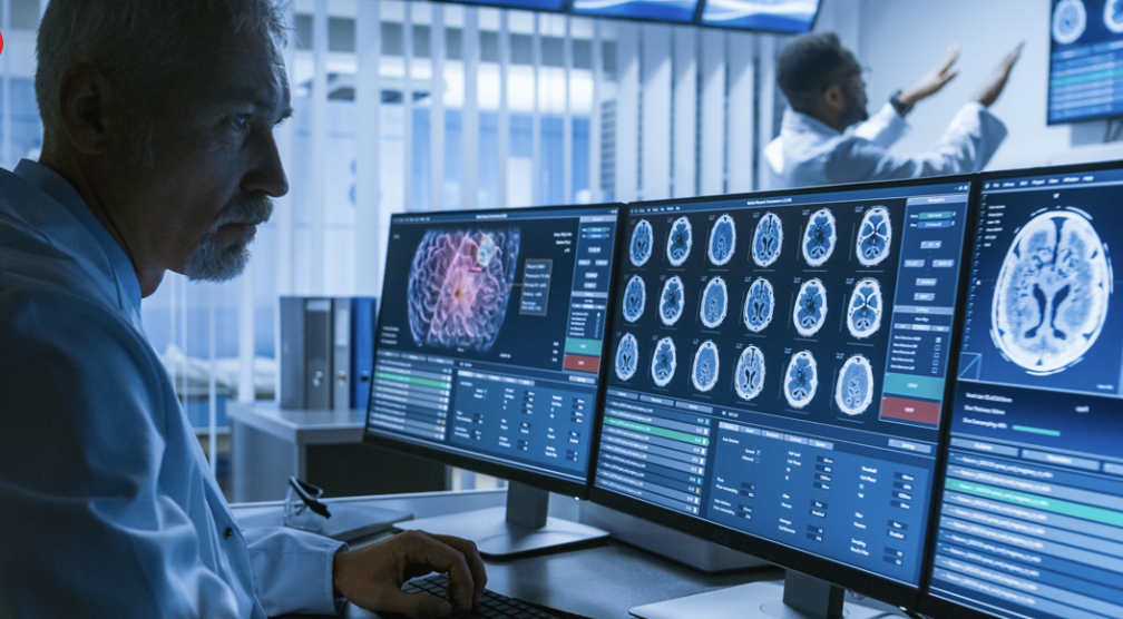
Where Imaging Analysis Falls Short
Radiologists who read CT scans are highly trained, and the interpretation of imaging in cancer surveillance is genuinely skilled work. But there are structural limits to what any individual reader can catch in a finite amount of time, reviewing one scan at one point in time.
Appendix cancer tends to recur along peritoneal surfaces in patterns that can be subtle early on. Small nodules, mild thickening, early mucinous change: these findings can be present on imaging and either not reported or reported as nonspecific, because they fall below the threshold of what looks definitively abnormal to a single reviewer.
This is one area where AI-assisted imaging analysis has genuine near-term potential. Rather than evaluating a single scan in isolation, machine learning models can be trained to compare images over time, tracking minute changes in tissue density, surface texture, and anatomical relationships across serial scans. They can also be trained on large datasets of confirmed recurrences, learning what early peritoneal disease tends to look like before it becomes obvious.
The goal is not to produce a diagnosis. It is to flag imaging findings that warrant closer attention, earlier in their development than a single-point review might catch them. For appendix cancer specifically, where the pattern of spread is often diffuse and gradual, that kind of longitudinal signal could meaningfully change the timeline of detection.

Circulating Tumor DNA and Its Limits
Liquid biopsy, particularly circulating tumor DNA (ctDNA) testing, has become an area of significant interest in cancer monitoring across multiple tumor types. The idea is straightforward: as tumors grow, they shed fragments of DNA into the bloodstream. Detecting those fragments can, in theory, indicate the presence of active disease before it is visible on imaging.
For appendix cancer patients, ctDNA remains a research-stage tool rather than a standard-of-care option, and its performance in this specific disease has not been well characterized in large studies. Some patients are accessing it through clinical contexts or ordering it independently, but interpreting the results without clear disease-specific reference data is genuinely difficult. A low-level signal may or may not be meaningful. A negative result does not rule out disease.
Where AI becomes relevant here is in interpretation over time rather than at a single point. Models trained on serial ctDNA measurements, correlated with imaging findings and clinical outcomes, can begin to identify what a subtle upward trend means for a specific patient profile, rather than requiring a clearly positive result to act on. That kind of longitudinal pattern analysis is not something a clinician can reliably do in their head across a large patient population, but it is exactly what machine learning is designed for.
This is still early, particularly for a rare cancer with limited datasets. But the trajectory is real, and it is worth watching.
| ASKING ABOUT CTDNA If you are interested in ctDNA testing as part of your monitoring, it is worth asking your oncologist whether there is a clinical context in which it would be interpretable for your situation. Results from ctDNA tests are most useful when there is a known mutation from your tumor to track. Without that anchor, results can be difficult to act on. |
The Case for Individualized AI Surveillance
One of the more significant shifts that AI could enable in cancer monitoring is moving away from fixed surveillance schedules toward monitoring that responds to what an individual patient’s data actually shows.
Current schedules, scan every three months for two years then every six months, are built around population-level risk curves. They represent a reasonable average, not a precise fit for any individual patient. A patient whose disease has been completely resected with clear margins and no elevated markers at any point during follow-up may be at lower near-term risk than someone whose initial surgery achieved only partial cytoreduction. A fixed schedule treats both the same way.
AI-assisted surveillance models could, in principle, adjust monitoring intensity based on a patient’s cumulative data: imaging trends, marker trajectories, time since treatment, molecular features of the original tumor, and comparison to outcomes in similar patients. Someone whose profile suggests lower risk might reasonably scan less frequently. Someone whose data shows early signals, even below the threshold of clinical concern, might be watched more closely.
For patients, this is not just a clinical question. More frequent scanning carries its own costs, including radiation exposure, anxiety, and financial burden. Less frequent scanning when it is clinically appropriate is not a reduction in care. It is better-calibrated care. And earlier intervention when something is actually shifting can change what treatment options are available.
Being Honest About Where Things Stand
Most of what is described in this post is not yet part of standard appendix cancer surveillance. AI-assisted imaging analysis is being studied but is not widely deployed. ctDNA interpretation in appendix cancer specifically lacks the disease-specific validation data needed to make it reliably actionable. Individualized surveillance models do not yet exist in a form that most oncologists can access for their patients.
The reason to pay attention to this now is not that it is available, but that it is directionally real. Imaging AI is advancing rapidly across oncology. Liquid biopsy is being integrated into cancer care in other tumor types and will eventually reach rare ones. Data sharing across institutions, which is the prerequisite for training models on appendix cancer specifically, is improving as registries and collaborative research networks grow.
For patients in a rare cancer community, staying informed about where the field is heading matters. It shapes the questions you ask, the research you follow, and, increasingly, the advocacy work that pushes these tools toward the diseases that need them most.
| Questions to Ask Your Doctor |
| • What does my specific monitoring schedule look like, and what is it based on? |
| • Which tumor markers are relevant for my subtype, and how should I interpret a normal result? |
| • Is there anything in my imaging history that has been noted as nonspecific or worth watching that we have not discussed in detail? |
| • Is ctDNA testing something that would be interpretable for my situation, given the known mutations in my tumor? |
| • At what point would a change in my markers or imaging prompt a change in my monitoring frequency or additional testing? |
| • Are there any clinical trials focused on surveillance or early recurrence detection in appendix cancer that I should know about? |
About This Series
This is the fifth post in Appendicure’s ongoing series on how artificial intelligence is beginning to intersect with appendix cancer. The final post will explore how AI could expand access to clinical trials for appendix cancer patients.
Read Part 1: Appendix Cancer and Surgical Decisions: How AI May Help Guide the Hardest Choice
Read Part 2: Why Appendix Cancer Is So Often Missed, and What Could Change That
Read Part 3: Not All Appendix Cancers Are the Same, and That Distinction Changes Everything
Read Part 4: Personalized Treatment in Appendix Cancer: Why One Plan Does Not Fit All
Leave a Reply