
Pseudomyxoma peritonei grade shapes almost every decision that follows a diagnosis, and it usually comes only after surgery. A new open-access study asks whether a routine CT scan, read with the help of AI, can estimate pseudomyxoma peritonei grade before the operation. The early answer is promising, and worth understanding, even though nothing here is ready for the clinic yet.
FROM OUR COMMUNITY. Many people in the Appendicure community learn their tumor grade only after surgery, once the pathologist has looked at the tissue. Grade shapes so much of what comes next, so the question of whether a scan could hint at it earlier is one a lot of us have wondered about. If you have thought about this, your experience belongs in the registry.
Pseudomyxoma peritonei grade is one of the most important words in an appendix cancer diagnosis. A low-grade tumor and a high-grade tumor can look similar on a report and behave in completely different ways. Grade helps guide how aggressive treatment should be, how closely someone is watched afterward, and how surgical teams plan an operation. The catch is that grade is usually confirmed by looking at tumor tissue under a microscope, which means waiting for a biopsy or for the surgery itself.
A study published in BMC Medical Imaging on July 28, 2025, tested a different starting point. The researchers, from Aerospace Center Hospital in Beijing, asked whether the information already sitting inside a preoperative CT scan could estimate grade on its own, before anyone picks up a scalpel.
What the study did
The team reviewed 158 patients with pathologically confirmed pseudomyxoma peritonei of appendiceal origin, treated between January 2015 and April 2024. Eighty-five had low-grade disease and 73 had high-grade disease. Every patient had a preoperative contrast-enhanced CT scan, and the researchers focused on the delayed phase, which is the set of images taken a while after the contrast dye is injected, when it has had time to settle into the tissue.
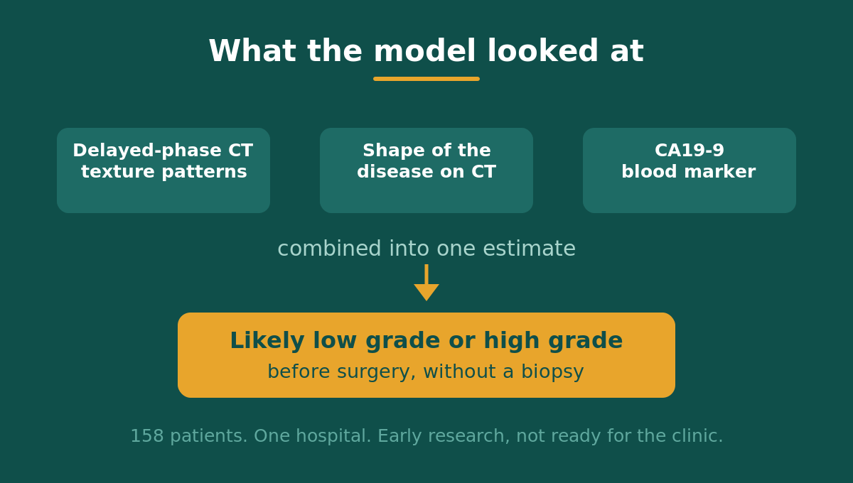
They used a technique called radiomics. Radiomics pulls hundreds of measurements out of a scan that the human eye cannot see, things like texture, density patterns, and the shape of the disease. Those measurements get fed into a computer model that learns which patterns line up with which grade. The team also folded in routine clinical data, including the blood markers CEA, CA19-9, and CA125, and the CT-based estimate of how much disease was present.
They built three versions of the model. One used only the clinical data. One used only the imaging. And one combined both. Then they tested how well each version told low-grade disease apart from high-grade disease.
What it found
The combined model did best at estimating pseudomyxoma peritonei grade. It reached an AUC of 0.91 when it was being trained and 0.88 when it was tested on cases it had not seen. AUC is a score from 0.5 to 1.0 that measures how well a test separates two groups, where 0.5 is a coin flip and 1.0 is perfect. A test score near 0.88 is considered strong. The combined model clearly beat the imaging-only and clinical-only versions, and it stayed well calibrated, meaning its confidence levels matched what actually happened.
The researchers also used a method called SHAP to see which features carried the most weight. The three that mattered most were two CT texture and shape measurements and the CA19-9 blood marker. The takeaway is that no single number did the work. The scan and the bloodwork together told a fuller story than either one alone.
KEY TAKEAWAY. A model built from a delayed-phase CT scan plus routine bloodwork estimated pseudomyxoma peritonei grade, low versus high, with an AUC of 0.88 on testing. That is a strong early result for a tool that needs no biopsy, though it has only been shown at one center so far.
A word about how this study defined pseudomyxoma peritonei grade
This study sorted patients into two buckets, low grade and high grade. That is a common shortcut in research, but it does not match how appendiceal disease is actually graded in careful practice. The standard-of-care framing that Appendicure follows, the 2025 Godfrey consensus guidelines, preserves a three-tier system. Moderately differentiated disease sits in the middle and should not be quietly folded into the high-grade group.
This matters for anyone reading a study like this. A model trained on a two-tier split of pseudomyxoma peritonei grade has never been asked to recognize that middle tier. So even if the numbers look strong, the tool as built cannot speak to where a moderately graded tumor would land. That is a real gap, not a small footnote, and it is worth keeping in mind before treating any two-tier result as the whole picture.
What this could mean for patients
The appeal here is timing. Knowing something about pseudomyxoma peritonei grade before surgery, rather than after, could help a care team plan. It might inform how extensive an operation to prepare for, how to counsel a patient about what to expect, or when a second opinion on the surgical plan makes sense. A scan-based estimate would never replace pathology, which remains the real answer. It would be an early clue, available at a point when there are no other clues.
It is also worth noting what this approach uses. A delayed-phase contrast CT and standard blood markers are things many patients already have on file. The study is not asking for exotic testing. It is trying to squeeze more information out of scans that are already being done.
What this study cannot tell us
This is early work. It comes from a single hospital, it looked back at past cases rather than following patients forward, and 158 patients is a small group for training and testing a model of this kind. The results have not been checked at other centers, and models like this often lose some accuracy when they meet scans from different machines and different radiology teams.
This is also not the first attempt at the same idea. An earlier study used a different CT feature, the pattern of disease coating the omentum, to estimate grade and reported similar accuracy. So this line of research is building, not breaking new ground. The honest way to read it is as one more careful step toward a tool that does not exist yet, rather than a finished test anyone can ask for today.
Most important, none of this changes care right now. There is no approved scan-based tool for pseudomyxoma peritonei grade in appendix cancer. Grade still comes from pathology, and treatment decisions still belong with an experienced multidisciplinary team.
Questions to bring to your team
These questions fit this study’s findings. Some may already be part of your conversations. Take what is useful.
- What is known about my pseudomyxoma peritonei grade right now, and how was it determined?
- Does my care team use a three-tier grading system, and where does my disease fall within it?
- Have my imaging and blood markers, like CEA, CA19-9, and CA125, been reviewed together as part of planning?
- Are any imaging-based or AI tools being used or studied at my center to help with surgical planning?
- How does my grade shape the plan for surgery and for follow-up afterward?
Why your data matters
A study like this is only possible because one hospital happened to have 158 scans and matched pathology in one place. Most appendix cancer patients are scattered across health systems that never compare notes. That is the exact gap the Patient-Led Global Appendix Cancer Registry is built to close. When patients pool their diagnosis, imaging history, treatment, and outcomes, researchers can ask better questions and test tools that estimate pseudomyxoma peritonei grade across many centers instead of just one. The registry now has IRB approval and an exempt determination, and your information is handled with care.
If you want your experience counted, you can add it here:
United States: https://form.jotform.com/261653563455159
International: https://form.jotform.com/261874541837063
ADD YOUR STORY. Joining the registry takes a few minutes and helps researchers study appendix cancer subtypes that are too rare for any single hospital to figure out alone.
Glossary
Pseudomyxoma peritonei (PMP). A condition where mucinous tumor spreads through the lining of the abdomen, most often from a tumor of the appendix.
Grade. A measure of how abnormal tumor cells look and how aggressively the disease tends to behave. Appendiceal disease is graded across three tiers, from low to high.
Delayed-phase CT. Images taken later in a contrast CT scan, after the dye has had time to move into the tissue.
Radiomics. A method that extracts many hidden measurements from a scan, such as texture and shape, and feeds them into a computer model.
AUC. A score from 0.5 to 1.0 for how well a test separates two groups. Higher is better, and 0.5 is no better than a guess.
SHAP. A way of showing which features a model leaned on most when it made a prediction.
Source
Wang Z, et al. A radiomics-based interpretable model integrating delayed-phase CT and clinical features for predicting the pathological grade of appendiceal pseudomyxoma peritonei. BMC Medical Imaging. 2025;25:300. Open access. https://doi.org/10.1186/s12880-025-01843-6
Keep reading: PET Scans and Appendix Cancer, Myth vs Fact and Watching for What Comes Back, How AI Could Change Recurrence Monitoring in Appendix Cancer.
Medical disclaimer. This post is for educational purposes only and is not medical advice. Research on appendix cancer evolves quickly. Treatment decisions should always be made with a qualified medical team familiar with your case. Appendicure is a 501(c)(3) nonprofit dedicated to appendiceal cancer patient education and advocacy and is not a medical provider.
Leave a Reply