

Appendix cancer imaging is improving, but we are still borrowing most of what we know from other cancers. The two advances most likely to help people with mucinous appendix cancer and pseudomyxoma peritonei soon are FAPI PET, a scan that finds tumors a standard PET misses, and diffusion MRI, which can map disease without radiation.
I read a lot of appendix cancer imaging research, because a scan is often the first place appendix cancer shows up and the main way we watch it over time. Most of that research is not built for our disease. It is built for colon, stomach, and ovarian cancer, and then borrowed for ours. When I dug into what actually has appendix data behind it, two things stood out as worth real attention. I want to walk through both in plain terms, and be honest about what they cannot do yet.
Why standard PET misses mucinous appendix cancer
A standard PET scan uses a radioactive sugar tracer. It lights up cells that burn a lot of sugar, which most cancers do. Mucinous appendix tumors do not. They are mostly mucus with relatively few tumor cells spread through it, and those cells use little of that sugar. So the tumor can stay dark on the scan even when disease is all over the belly. In mucinous appendix cancer, a standard PET picks up disease only about one time in three. I wrote more about this in PET Scans and Appendix Cancer, Myth vs Fact. The short version is that a clean PET does not mean you are clear.
That is the problem the first advance is built to solve.
FAPI PET, a scan that targets the tumor’s scaffolding
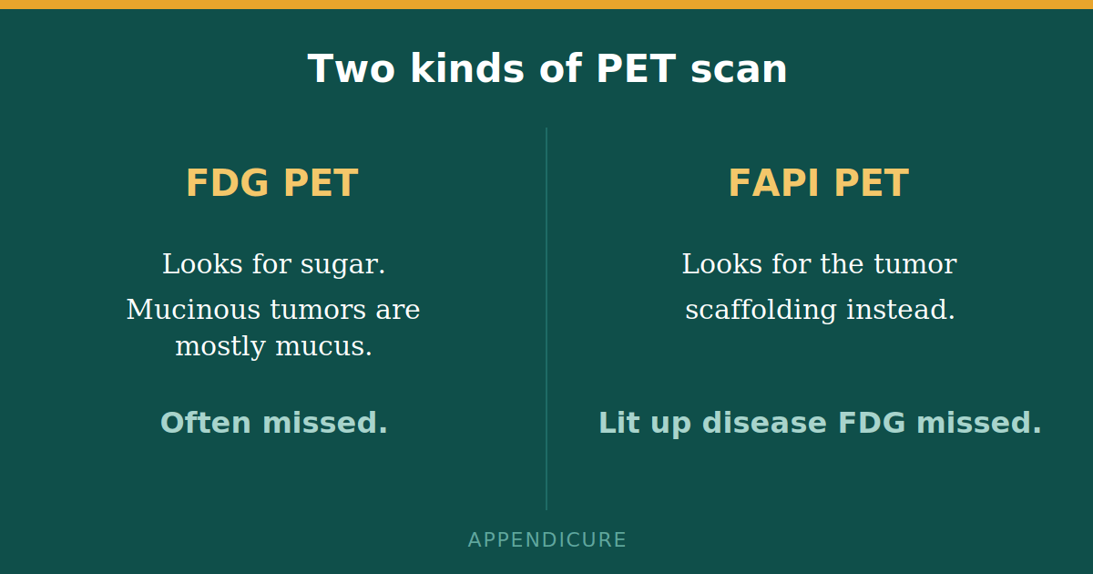
FAPI is a newer tracer. Instead of chasing sugar, it sticks to a protein found on the support cells that surround a tumor, the scaffolding that mucinous tumors are full of. So the tumor can light up even when it is mostly mucus.
There is now one study in appendix patients specifically. Researchers scanned 22 people with appendiceal tumors and compared FAPI PET against standard PET and a CT scan. In this study, FAPI detected the primary tumor in 87.5 percent of patients, compared with 43.8 percent for standard PET. It also identified peritoneal disease in every patient who had it, and it picked up more than twice as many spots of disease as standard PET, 108 areas against 43. In one published case, FAPI showed widespread disease that a standard PET had shown as completely clean.
I want to be careful here. This is one small study from a single center, read without a tissue check to confirm the target, and FAPI scans are not widely available or routinely covered yet. This is early, promising evidence, not proven or standard. But it points at exactly the gap our community keeps falling into, and it is the molecular scan I am watching most closely.
Better MRI, and why it matters for surveillance
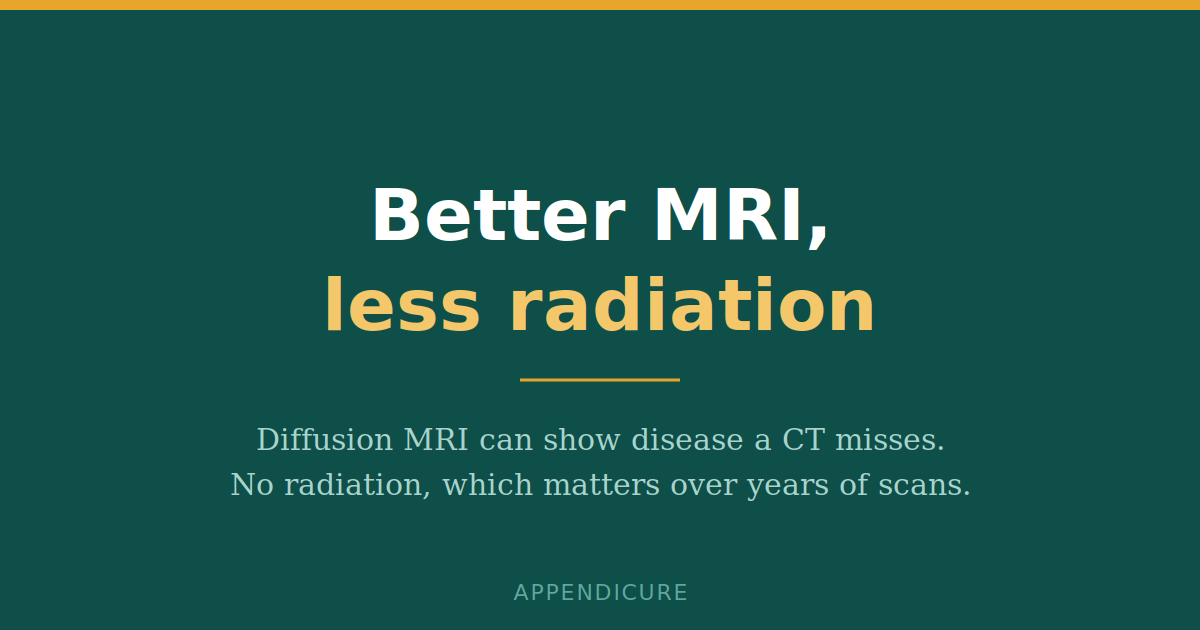
The second advance is diffusion MRI. A regular scan shows anatomy. It measures how water moves inside tissue, and tumor traps water, so disease stands out. Paired with contrast MRI, it does two useful things for mucinous disease.
First, it maps how much disease is present before surgery. In an appendix-heavy group of patients, diffusion MRI combined with contrast MRI correctly estimated disease burden in roughly 88 percent of patients. Second, the way the tissue takes up contrast can hint at whether mucinous disease is low grade or high grade, without another biopsy. And MRI uses no radiation.
That last part matters more than it sounds. Most of us live in surveillance, getting scanned again and again for years. Today that surveillance runs mostly on CT, paired with the CEA and CA19-9 blood markers. CT is still the backbone. But radiologists increasingly reach for MRI to cut radiation, especially in younger patients facing a decade or more of follow-up. Whole-body MRI is being studied for the same reason, primarily in colorectal and other abdominal cancers. The catch is that the strongest whole-body MRI numbers come from colon cancer, not ours, and mucinous disease can be faint on diffusion scans, so it may not carry over cleanly. That question is still open.
One thing every patient should know about scans and surgery
CT scans frequently underestimate how much disease is actually present. In studies of appendix and PMP patients, the amount of disease a CT shows is regularly less than what the surgeon actually finds. A scan that looks bad is not by itself a reason to be told surgery is off the table. If you are ever told your disease is too widespread to operate on, that is a moment to get a second opinion from a peritoneal surface specialist who does cytoreductive surgery. The scan is a guide, not the final word.
Where appendix cancer imaging is headed
Most of what gets billed as the future of imaging is not really about appendix cancer imaging yet. Photon-counting CT, PET combined with MRI, dual-energy CT, fluorescent dyes that make tumor glow during surgery, and software that predicts biology from a scan are all advancing quickly, but nearly all of the evidence comes from other cancers. For appendix disease specifically, they are ideas to watch, not tools to ask for. The same is true for most artificial intelligence tools. I covered two of the furthest-along AI ideas already, in reading PMP grade from a CT scan and in how AI could change recurrence monitoring.
Two honest limits belong on all of this. Nearly every appendix study here is small and from a single center, so the numbers may shift as more people are studied. And this evidence is about mucinous appendiceal tumors and PMP. Goblet cell adenocarcinoma, signet ring cell carcinoma, and neuroendocrine tumors of the appendix behave differently on scans, and the FAPI and MRI findings above should not be assumed to apply to them. The current standard-of-care reference for our disease is the 2025 PSM Consortium consensus, not the colon cancer playbook.
What I am doing about the gap
The theme running through all of this is the same. Our disease keeps getting scanned with tools calibrated for other cancers, and the studies that would fix that do not exist. There is no large, dedicated, multi-institution imaging effort focused specifically on appendix cancer. That is exactly the kind of gap a patient community can help close, by pooling what individual hospitals never see enough of on their own. It is why I keep pushing on research partnerships, and why the work Appendicure does depends on patients raising their hands.
If you want to help that work move faster, you can support Appendicure here.
Add your data to the Patient-Led Global Appendix Cancer Registry. The registry collects molecular, genomic, and pathology information from appendix cancer patients so researchers can study our disease directly instead of borrowing from others. It takes a few minutes and it helps every subtype.
Join the Registry: United States Join the Registry: InternationalQuestions patients ask about appendix cancer imaging
Can an MRI find appendix cancer that a CT scan misses?
Sometimes, yes. Diffusion MRI measures water movement in tissue and can show small mucinous deposits that a CT scan does not pick up. It also uses no radiation, which is why it is being used more for long-term surveillance.
What is FAPI PET?
FAPI PET is a newer scan that targets the support cells around a tumor instead of the tumor’s sugar use. Because mucinous appendix tumors are mostly mucus with few active cells, they often hide on a standard PET but can light up on a FAPI PET. It is promising but still early, and not widely available yet.
Why do PET scans miss appendix cancer?
Standard PET looks for cells that burn a lot of sugar. Mucinous appendix tumors burn very little, so they can stay dark even when disease is widespread. A clean PET does not rule out disease.
Does a bad-looking scan mean surgery is off the table?
Not by itself. Scans tend to undercount how much disease is really there. If you are told surgery is not possible, get a second opinion from a peritoneal surface specialist before accepting that.
Is appendix cancer imaging getting better?
Yes, but unevenly. Appendix cancer imaging is improving fastest in two places, FAPI PET and diffusion MRI, while most other advances are still borrowed from other cancers and not yet proven for our disease.
Read more
PET Scans and Appendix Cancer, Myth vs Fact
Reading Pseudomyxoma Peritonei Grade From a CT Scan
Watching for What Comes Back, How AI Could Change Recurrence Monitoring
Sources: FAPI in appendiceal neoplasms, Cancer Imaging 2024. Diffusion and contrast MRI for peritoneal cancer index, Annals of Surgical Oncology 2012, and mucinous appendiceal MR staging, AJR 2008. CT undercounting of surgical disease, Journal of Gastrointestinal Surgery 2016. Surveillance imaging review, British Journal of Radiology 2024. Standard of care, PSM Consortium consensus 2025. Full cited research dossier available from Appendicure.


Leave a Reply