

Appendix cancer biomarker testing usually means several separate tests, and they do not always agree with each other. A case report posted in August 2026 describes a young woman whose MMR protein test and MSI test gave opposite answers, and what her doctors did once they stopped treating one of those results as the whole picture.
A 25-year-old woman in Japan went to a clinic with a swollen abdomen. She had been having abdominal pain for months. Scans showed masses on both ovaries, nodules scattered across her peritoneum, and a large amount of fluid. She was referred to gynecology with suspected ovarian cancer.
A colonoscopy and a set of stains on her biopsy pointed somewhere else. The tumor had started in her appendix. It was signet ring cell adenocarcinoma, and it had spread to both ovaries and to her cervix.
What appendix cancer biomarker testing actually measures
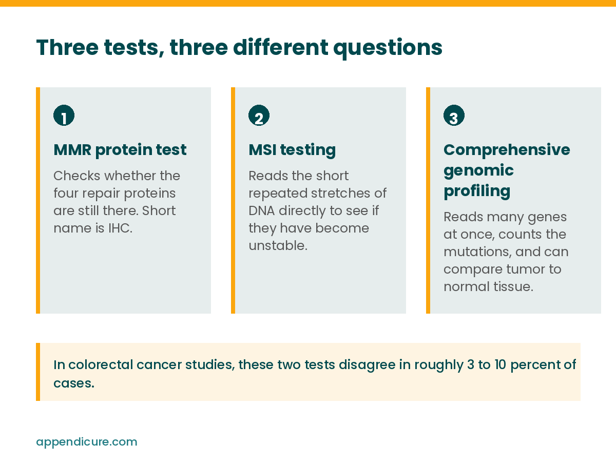
Appendix cancer biomarker testing is not one test. Three separate ones show up in this story, and each asks a different question about the tumor.
The MMR protein test checks whether four specific repair proteins are still present in the tumor tissue. Pathologists call it immunohistochemistry, or IHC. When one or more of those proteins is missing, the report says mismatch repair deficient, usually written dMMR.
MSI testing skips the proteins and reads the DNA. It looks at short repeated stretches called microsatellites to see whether they have become unstable. A stable result is written MSS. An unstable one is MSI-high.
Comprehensive genomic profiling, or CGP, reads many genes at once. It can report MSI status, count how many mutations the tumor is carrying, which is called tumor mutational burden or TMB, and check individual genes like RAS, BRAF, and HER2. When it is run on tumor tissue and normal tissue side by side, the comparison can also flag a change that looks inherited rather than something the tumor developed on its own. That is a flag rather than a final answer, and confirming it usually means a separate germline test through a genetics service.
What happened in this case
Her MMR protein test showed loss of MLH1 and PMS2. MSH2 and MSH6 were still there. On that result by itself, her tumor was mismatch repair deficient, which pointed her team toward immunotherapy.
Then the MSI test came back microsatellite stable. The genomic profiling agreed and put her tumor mutational burden at 3.7 mutations per megabase, which is low. Both of those results point away from immunotherapy.
Her team followed the MMR result and started nivolumab plus ipilimumab. After three cycles her scans showed the cancer had grown, and her fluid had built up enough to need draining again.
They switched to mFOLFOX6 chemotherapy. After two cycles the fluid still had not come down.
At that point they went back to the full genomic profile and read it as a whole. Her tumor was RAS and BRAF V600E wild type, HER2 negative, and carried none of the major changes known to block anti-EGFR drugs. Her team added panitumumab, an anti-EGFR antibody, to the mFOLFOX6. Her fluid nearly disappeared, her abdominal nodules shrank, and the response was scored as a partial response. By the time the authors wrote the case up, she had received ten more cycles and was still under control.
Why two tests of the same system can disagree
Published colorectal cancer studies put the disagreement rate between MMR protein testing and molecular MSI testing at roughly 3 to 10 percent. Comparable figures for appendiceal cancer specifically have not been established.
Confirmation testing catches some of it. In the CheckMate 8HW colorectal cancer trial, 303 patients entered the first-line comparison based on MSI-high or dMMR testing done at their own hospital. Central testing confirmed that status in 255 of them. The trial does not break down why the rest were not confirmed, so those are not all wrong local results, but the gap is a reason to take a second look when a result does not fit the rest of the picture.
Several things can cause it. Not enough tumor in the sample. Handling problems before the test is run. Real differences between tumor cells in one part of the tissue and another. Differences in how sensitive each assay is.
In her case the authors ruled some of those out. She had not had chemotherapy before either test. The protein loss was even across the tissue rather than patchy. The genomic profiling found no mutation in MLH1 itself. They offer methylation of the MLH1 promoter as one possible explanation, though they did not test for it. They raise a simpler possibility too, which is that losing the protein did not damage the repair system enough to produce a hypermutated tumor.
That first explanation has a wrinkle worth naming. An MD Anderson study of 108 appendiceal carcinomas concluded that MLH1 promoter methylation does not appear to be a mechanism for microsatellite instability in the appendix, unlike in the right colon where it is the usual cause. The hypothesis these authors offer is reasonable for her tumor, but it runs against what the larger appendiceal series found.
The inherited finding nobody was looking for
Her genomic profiling was run on tumor and normal tissue together. That paired setup found a TP53 mutation, p.R248Q, sitting in her normal tissue at a variant allele frequency of 46.8 percent, which means she was born with it. ClinVar classifies it as pathogenic. It confirmed Li-Fraumeni syndrome, an inherited condition that raises the risk of several cancers.
Both of her parents were tested for the same variant and neither one carried it, so it appears to have arisen new in her. No family history would have flagged it. Nothing about how she presented would have prompted the test on its own. It surfaced from a panel that was run to help pick a chemotherapy.
I wrote the plainer version of what inherited testing adds, and why it reaches your relatives, in appendix cancer genetic testing.
What this case does not show
This is one patient. The authors say so plainly and list four limits of their own.
She received panitumumab on top of mFOLFOX6, so there is no way to separate what the panitumumab did from what the chemotherapy did.
Anti-EGFR evidence in appendiceal cancer is thin. The idea of ruling out resistance mutations before choosing an anti-EGFR drug was developed in metastatic colorectal cancer, not in this disease.
The reason her two tests disagreed was never established.
And this is signet ring cell appendiceal adenocarcinoma, a subtype that makes up roughly 4 to 5 percent of primary appendiceal carcinomas. Nothing in this case covers LAMN, HAMN, goblet cell adenocarcinoma, mucinous adenocarcinoma, appendiceal neuroendocrine tumors, or pseudomyxoma peritonei.
One more thing about the source. This is a preprint posted on Research Square on August 28, 2026, and it is marked under review. It has not been through peer review. I am writing about it because the testing question sitting underneath it is one I see members hit right now, not because a single case changes anyone’s treatment.
What to ask your care team about appendix cancer biomarker testing
Has my tumor had MMR protein testing, MSI testing, and tumor mutational burden measured, or only some of those? If all of them were done, did the results agree with each other? Has comprehensive genomic profiling been run, and was it run on tumor tissue alone or on tumor and normal tissue together?
That last question decides whether an inherited finding can be spotted at all. A tumor-only panel cannot tell you what you were born with.
Questions people ask
Can a tumor be mismatch repair deficient and microsatellite stable at the same time?
Yes. Published colorectal cancer studies put the disagreement between MMR protein testing and MSI testing at roughly 3 to 10 percent. The two tests measure different things, so one can come back abnormal while the other comes back normal. Equivalent figures for appendiceal cancer have not been established.
Does a dMMR result always mean immunotherapy will work?
No. In this case report the tumor was dMMR on protein testing but microsatellite stable with a low tumor mutational burden, and nivolumab plus ipilimumab did not control it. Treatment decisions belong to you and your oncologist, and one case does not set a rule.
What is the difference between genetic testing and genomic profiling?
Genomic profiling reads the tumor. Genetic testing, also called germline testing, reads what you were born with. Some paired tumor and normal panels can flag both at once, which is how the Li-Fraumeni syndrome in this case surfaced. An inherited finding that turns up on a tumor panel still usually needs to be confirmed and handled through a genetics service.
Does appendix cancer biomarker testing require a new biopsy?
Often not. These tests usually run on tumor tissue that has already been removed and stored. Ask your team whether your existing tissue can be used.
Read more
MSI Testing in Appendix Cancer: The Rare 3% That Matters
Appendix Cancer Genetic Testing: Why Dr. Shen Suggested a Germline Test for David
Appendix Cancer Mutation Testing: Know What Your Tumor Is Made Of
Wild Type Appendix Cancer: What That 1 Word on Your Report Actually Means
Add your testing results to the registry
Nobody knows how often appendix cancer biomarker testing comes back with results that disagree. Appendiceal-specific data on that question is very limited. The Patient-Led Global Appendix Cancer Registry gathers mutation, biomarker, and pathology data from patients directly. It takes about ten minutes. The enrollment links are region-specific, so please use the one that matches where you live.
Join the Registry: United States Join the Registry: International
Already enrolled and need to add a new report or fix an earlier entry? Use the registry update form. Appendicure runs on donations, and if you want to help fund this work you can give here.
Sources
Ando T, Tajiri Y, Noda Y, et al. Comprehensive genomic profiling-guided treatment selection in appendiceal adenocarcinoma with discordant mismatch repair findings: a case report and literature review. Research Square preprint, posted August 28, 2026. Not peer reviewed. https://doi.org/10.21203/rs.3.rs-10434247/v1
Evrard C, Tachon G, Randrian V, Karayan-Tapon L, Tougeron D. Microsatellite instability: diagnosis, heterogeneity, discordance, and clinical impact in colorectal cancer. Cancers. 2019;11:1567. https://doi.org/10.3390/cancers11101567
Andre T, Elez E, Van Cutsem E, et al. Nivolumab plus ipilimumab in microsatellite-instability-high metastatic colorectal cancer. New England Journal of Medicine. 2024;391(21):2014-2026. https://doi.org/10.1056/NEJMoa2402141
Taggart MW, Galbincea J, Mansfield PF, et al. High-level microsatellite instability in appendiceal carcinomas. American Journal of Surgical Pathology. 2013;37(8):1192-1200. https://doi.org/10.1097/PAS.0b013e318282649b
McCusker ME, Cote TR, Clegg LX, Sobin LH. Primary malignant neoplasms of the appendix: a population-based study from the Surveillance, Epidemiology and End Results program, 1973 to 1998. Cancer. 2002;94:3307-3312. https://doi.org/10.1002/cncr.10589
This post is patient education from Appendicure and is not medical advice. Testing and treatment decisions should be made with your own care team.
Leave a Reply