


A preprint from The University of Manchester and The Christie, posted October 28, 2026, found higher levels of collagen XII, a protein encoded by the COL12A1 gene, in the support tissue around appendiceal mucinous tumors and PMP than in tissue without tumor involvement. In a lab model, fibroblasts grown from PMP increased how much PMP tumor cells spread into a gel, and turning down the COL12A1 gene in those fibroblasts reduced the tumor cells’ spread. The tissue studied included both low-grade and high-grade disease. The paper has not undergone formal journal peer review, and it did not test a treatment.
Most of what patients hear about PMP centers on the mucin and the tumor cells. A pathology report describes those cells: their grade, how they look under the microscope, and where they’ve spread. Much less gets said about the tissue around them. That support tissue is called the stroma. It includes fibroblasts, immune cells, blood vessels and the extracellular matrix, a mesh of proteins such as collagen that holds tissue together. A new study from Manchester focused on two parts of it, the fibroblasts and the matrix proteins.
Who did the study
Dr. Milly McAllister, a postdoctoral researcher at The University of Manchester, and Prof. Patrick Caswell, Professor of Cancer Cell Biology at the university, led the work. Prof. Omer Aziz, who leads the Peritoneal Tumour Service at The Christie in Manchester, is also an author. The researchers studied tissue collected during surgery from nine patients.
The paper was posted on bioRxiv, a site where researchers share work before it appears in a journal. It has not undergone formal journal peer review, and the findings could change.
Before writing about it, I emailed Prof. Aziz with three questions. He introduced me to Dr. McAllister and Prof. Caswell. Prof. Caswell answered my questions and sent an updated patient pathology table.
What the team measured
The team first measured which proteins were present in the tissue. They compared appendiceal mucinous tumors with uninvolved appendix, and PMP in the peritoneum, the lining of the abdominal cavity, with uninvolved peritoneum. Uninvolved means tissue without tumor involvement. It came from surgical patients, not from healthy volunteers.
Both kinds of tumor tissue showed changes in the matrix. There were more mucins, more of the matrix proteins that form long fibers, and more of the proteins that control how collagen is arranged. Collagen XII was raised in both the appendix tumors and the peritoneal disease.
The team also used tissue mapping, a method that shows where selected proteins are located, to see where collagen XII sat. Collagen XII levels were higher in the stroma of appendix tumors and peritoneal disease than in the corresponding uninvolved tissue. A separate analysis found more collagen and greater alignment in peritoneal tumors than in uninvolved peritoneum. The peritoneal tumors also had a higher proportion of dense matrix than the appendix tumors. These findings suggest that the collagen network had been remodeled.
I asked whether the protein comparisons were corrected for the large number of proteins tested at once. Prof. Caswell told me the p-values were adjusted.
What happened in the lab model
To see whether the matrix affects how PMP cells behave, the team grew a PMP cell line called N14A as tiny balls of cells inside a collagen gel. Then they measured how much the tumor cells spread into the gel. In this assay, adding fibronectin, another matrix protein, increased tumor-cell invasion, while adding biglycan reduced it.
Next they added fibroblasts grown from the peritoneal disease of two patients. In this model, the fibroblasts increased how much the PMP cells spread into the gel. When the team turned down the COL12A1 gene in the fibroblasts, the tumor cells spread less.
One of my questions was whether the invasion measurement counted only the tumor cells or also fibroblasts moving into the gel. Prof. Caswell told me it counts only the tumor cells. His team labels the two cell types with different fluorescent dyes so they can be told apart, and he said they’ll make that clearer in the manuscript.
Why the low-grade tissue mattered to me
Most people I talk to have low-grade disease, and they rarely see lab work that includes their tissue. The table published with the preprint showed mostly high-grade peritoneal samples in the protein analysis, so I asked whether the finding would hold in low-grade disease.
Prof. Caswell sent an updated table and said the information could be shared. He explained that his team often receives samples and starts its analysis before the detailed pathology is available. The tissue has to be processed quickly to preserve it, and the anonymized pathology report takes time to arrive.
In the protein analysis, two of the three appendix tumors were low-grade appendiceal mucinous neoplasms (LAMN) and one was a high-grade appendiceal mucinous neoplasm (HAMN). Of the four peritoneal samples, one was low-grade and three were high-grade. In the tissue mapping, three of the five appendix tumors were LAMN and two were HAMN, and one of the five peritoneal samples was low-grade. Many of the same patients contributed to both analyses. Of the two fibroblast cultures, one came from a patient with low-grade peritoneal disease and one from a patient with high-grade disease.
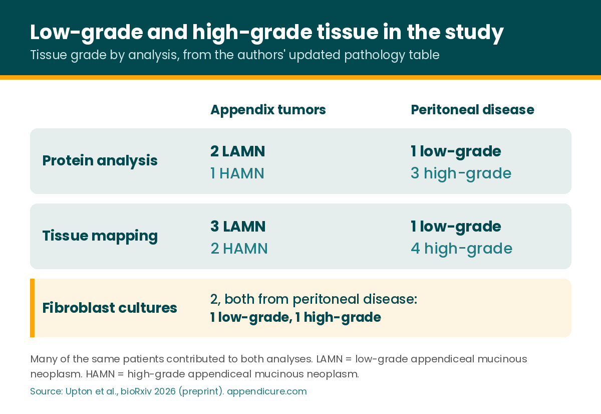
The grade of a patient’s appendix tumor and the grade of their peritoneal disease aren’t always the same, and the updated table includes an example of that.
Prof. Caswell told me the team believes the increase in collagen XII occurs in both low-grade and high-grade disease. Most appendix tumor samples were low-grade, although the total number was small. The proteomics and tissue-mapping analyses each included peritoneal disease from only one low-grade patient. “An important follow up would be to specifically analyse COL12A1 levels in a larger cohort of patient primary and disseminated disease,” said Prof. Patrick Caswell, Professor of Cancer Cell Biology and Head of the Division of Cell Matrix Biology and Regenerative Medicine at The University of Manchester. “A well documented and annotated cohort is one of the things [Prof. Omer Aziz] and the whole team has been building over the years and we hope to be able to make the most of this as soon as possible.”
What this study does not show
No experimental treatment was given to patients as part of this study, and no drug was tested. The lab experiments used one PMP cell line and fibroblasts from two patients. The tissue came from nine patients. Examining several areas from one patient provides more detail, but it does not increase the number of patients studied. These findings should not be generalized to other appendix cancer subtypes.
This study provides no evidence to change current treatment. For background on how appendiceal tumors are managed, the 2025 consensus guidelines from the Peritoneal Surface Malignancies Consortium lay out current expert recommendations.
How this connects to earlier work
In July I wrote about a Moffitt Cancer Center study that compared primary appendix tumors with peritoneal metastases from 14 patients. It measured gene activity separately in the tumor cells and in the surrounding tissue. Within the tumor cells, the peritoneal metastases showed more activity in collagen metabolism genes than the primary tumors. In the surrounding tissue, both primary and metastatic tumors showed more activity in protein-making pathways than normal appendix.
The Manchester study measured proteins and looked at collagen XII in the stroma. Both studies point to collagen biology as an area for further research, although they examined different aspects of it.
Where pathology reports fit
A routine pathology report generally doesn’t measure collagen XII levels. A report does show grade and subtype, which is the information Prof. Caswell’s team had to wait for. Accurate, complete reports help researchers describe who is in a study and plan future studies with enough low-grade patients in them.
Add your pathology report to the registry
The Appendicure Patient-Led Global Appendix Cancer Registry collects pathology, molecular and genomic reports from appendix cancer and PMP patients around the world. It’s IRB-reviewed.
Already enrolled? Add a new report or update your record here.
Questions about collagen XII and this study
What is the stroma?
The stroma is the support tissue around tumor cells. It includes fibroblasts, immune cells, blood vessels and the extracellular matrix. This study focused on fibroblasts and matrix proteins around appendiceal mucinous tumors and PMP.
What’s the difference between COL12A1 and collagen XII?
COL12A1 is the gene. Collagen XII is the protein it makes. The study measured collagen XII in tissue and turned down the COL12A1 gene in fibroblasts in the lab.
Did the study include low-grade disease?
Yes. Most of the appendix tumor samples were LAMN, and one of the two fibroblast cultures came from low-grade peritoneal disease. The proteomics and tissue-mapping analyses each included peritoneal disease from only one low-grade patient.
Did the researchers test a drug?
No. In a lab model, turning down the COL12A1 gene in fibroblasts reduced how much PMP cells spread into a gel. No drug was tested.
Does this change my treatment?
No. It’s laboratory research on tissue from nine patients, and it provides no evidence to change current treatment.
Read more
Appendix Cancer Tumor Microenvironment: It’s Not Just the Cancer Cells
Why PMP Behaves the Way It Does: A New Look at the Biology of the Disease
Your Tumor Is More Than Its Mutations: What New Appendix Cancer Research Is Showing Us
Sources
Upton M, Nagaraju R, Castaneda A, et al. Stromal COL12A1 defines a pro-invasive extracellular matrix niche in appendiceal mucinous neoplasms and pseudomyxoma peritonei. bioRxiv. Posted October 28, 2026. doi:10.64898/2026.10.26.761946. Preprint, not peer reviewed.
Updated patient pathology table provided by Prof. Patrick Caswell, November 4, 2026.
Park MA, Jacobson R, Genilo-Delgado M, et al. The Transcriptomic Landscapes of Appendiceal Primary and Metastatic Tumors are Distinct. Ann Surg Oncol. 2025;32(5):3660-3671. doi:10.1245/s10434-025-16939-0
Godfrey EL, et al; Peritoneal Surface Malignancies Consortium Group. Consensus Guideline for the Management of Patients with Appendiceal Tumors, Part 1: Appendiceal Tumors Without Peritoneal Involvement. Cancer. 2025. doi:10.1002/cncr.35867
Godfrey EL, et al; Peritoneal Surface Malignancies Consortium Group. Consensus Guideline for the Management of Patients with Appendiceal Tumors, Part 2: Appendiceal Tumors With Peritoneal Involvement. Cancer. 2025. doi:10.1002/cncr.35874
Appendicure is a patient-led nonprofit. If you’d like to support its work, you can donate here.
This post is patient education, not medical advice.
Leave a Reply