Yes, an appendix abscess can be associated with an underlying appendiceal tumor. A 2025 prospective study of 370 patients with an abscess around the appendix found tumors in 14.3 percent, compared with 1.5 percent of patients with uncomplicated appendicitis. Nearly all of those tumors were in patients over 35. New imaging research published on August 28, 2026 lays out six warning signs that should prompt a closer look.
A lot of people in our community started the same way. Bad stomach pain. A trip to the emergency room. A scan that showed a pocket of infection near the appendix. Antibiotics, maybe a drain, and a plan to follow up later. Nobody said the word cancer, because nothing on that first scan said cancer.
Months later, sometimes years later, the real answer showed up.
A team of radiologists in Italy just published a paper about exactly this problem. They call it the “cancer-abscess,” and they are careful to say it is not a new disease. It is a teaching tool. It is their way of telling other radiologists to slow down when a collection of fluid does not behave the way an infection should.
What an appendix abscess can hide
The paper describes two patients. The first one matters most to us.
That patient had an appendectomy. About a month later they came back with belly pain and a fever, which is a normal thing to happen after abdominal surgery. An ultrasound showed what looked like a 7 cm pocket of fluid near the cecum, right where you would expect a collection to form after surgery.
Two things did not fit. There were swollen lymph nodes nearby, some almost 2 cm across. There were similar nodes up under the liver. That is not how a simple post-surgical infection behaves, so the team ordered a CT scan with contrast.
The CT showed something very different. There was a mass at the old appendectomy site, roughly 6 by 4 by 6 cm. Part of it was fluid and dead tissue, which is what an abscess looks like. But part of it was solid tissue that lit up brightly with contrast dye. Contrast can light up inflamed tissue too, so that finding does not prove cancer on its own. It does mean there is living tissue in there that an ordinary pocket of infection would not have. There were also nodules scattered in the fat around the organs, along the mesentery, and down the right side of the abdomen.
They scanned again later. The mass had not changed despite treatment. That is not proof of anything either, because abscesses can be stubborn. It was one more reason to keep asking.
The workup confirmed what the imaging had raised concern about. This was not simply a routine collection after surgery. There was a tumor involved.
Six things that should trigger a second look
This is the part of the paper I would want any patient or family member to have. The authors list the imaging features that should make a radiologist question an infection diagnosis.
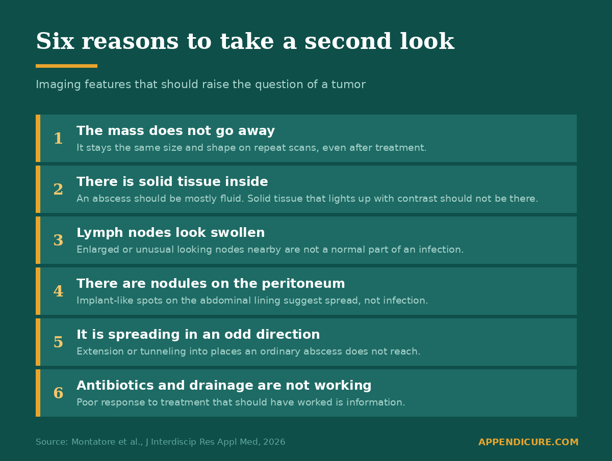
None of these prove anything on their own. A radiologist can see one of them and still be looking at an ordinary infection. What the authors are arguing is that these findings should stop the story from ending at “abscess,” and should trigger more imaging, a conversation across specialties, and sometimes a biopsy or surgery.
How often is there actually a tumor under an appendix abscess
The imaging paper does not answer that. It is two patients. It is a teaching case, not a study of how common this is, and the authors say so plainly.
Other research does answer it, and the numbers are higher than most people expect.
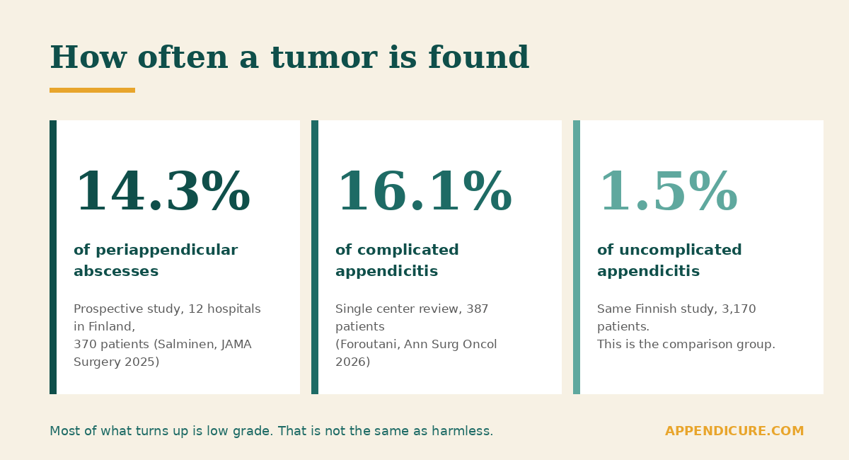
The strongest study is from Finland. Researchers followed more than 6,000 people with appendicitis across twelve hospitals. Of those, 396 had an abscess around the appendix. In the 370 who had tissue examined, 14.3 percent turned out to have a tumor. In people with straightforward appendicitis and no abscess, the rate was 1.5 percent.
Age was the one thing that predicted it. Almost every tumor was found in someone over 35. The Finnish team concluded that, at minimum, patients over 35 who are treated without surgery for an appendix abscess should go on to have an interval appendectomy, meaning the appendix comes out a few months later so it can be examined.
A separate review at the University of California, San Francisco, published in January 2026, looked at 387 patients whose scans showed complicated appendicitis. That group includes perforation, abscess, phlegmon, gangrene, and mucocele. They found tumors in 16.1 percent. The rate changed a lot depending on what the scan showed. When there was a mucocele it was 58 percent. With an abscess it was 18 percent. With a phlegmon, 16 percent. With a perforated appendix, 12 percent.
Their list of predictors overlaps almost exactly with the imaging paper: older age, a bigger appendix, swollen lymph nodes, enhancement of the peritoneum, and the absence of the fat streaking you would expect with a plain infection.
Where researchers disagree
I want to be straight about this, because there is a real argument happening in the journals right now and I do not think patients are served by hearing only one side.
A study out of Paris published in JAMA Surgery in 2026 looked at 2,293 appendectomies. They found tumors in 1.6 percent, and most of them were small, low-grade neuroendocrine tumors under 1 cm that never came back. Aggressive cancers were rare. Their argument is narrower than it first sounds. They found that antibiotic-only treatment looks safe from a cancer standpoint for carefully selected patients, meaning people without the high-risk features on their history and their scan. That is not the same as saying it is safe for everyone. A separate American study across six hospitals found incidental tumors in 1.3 percent, and did not find that complicated disease raised the odds, though it also excluded anyone whose scan already suggested a mass.
The Paris group wrote a letter to the editor on July 20 of this year challenging the San Francisco findings. The San Francisco group published its reply on August 3. A surgical oncology team in Pittsburgh followed with an editorial calling for the whole management algorithm to be rewritten. This is being worked out in public, in the space of about six weeks, and it is not settled.
The disagreement mostly comes down to who you are counting. If you count everyone who walks in with appendicitis, tumors are uncommon. If you count only the people whose scan shows an appendix abscess or a mucocele, the rate jumps by roughly ten times. Both things are true. They are different groups of people.
Why this matters for our community specifically
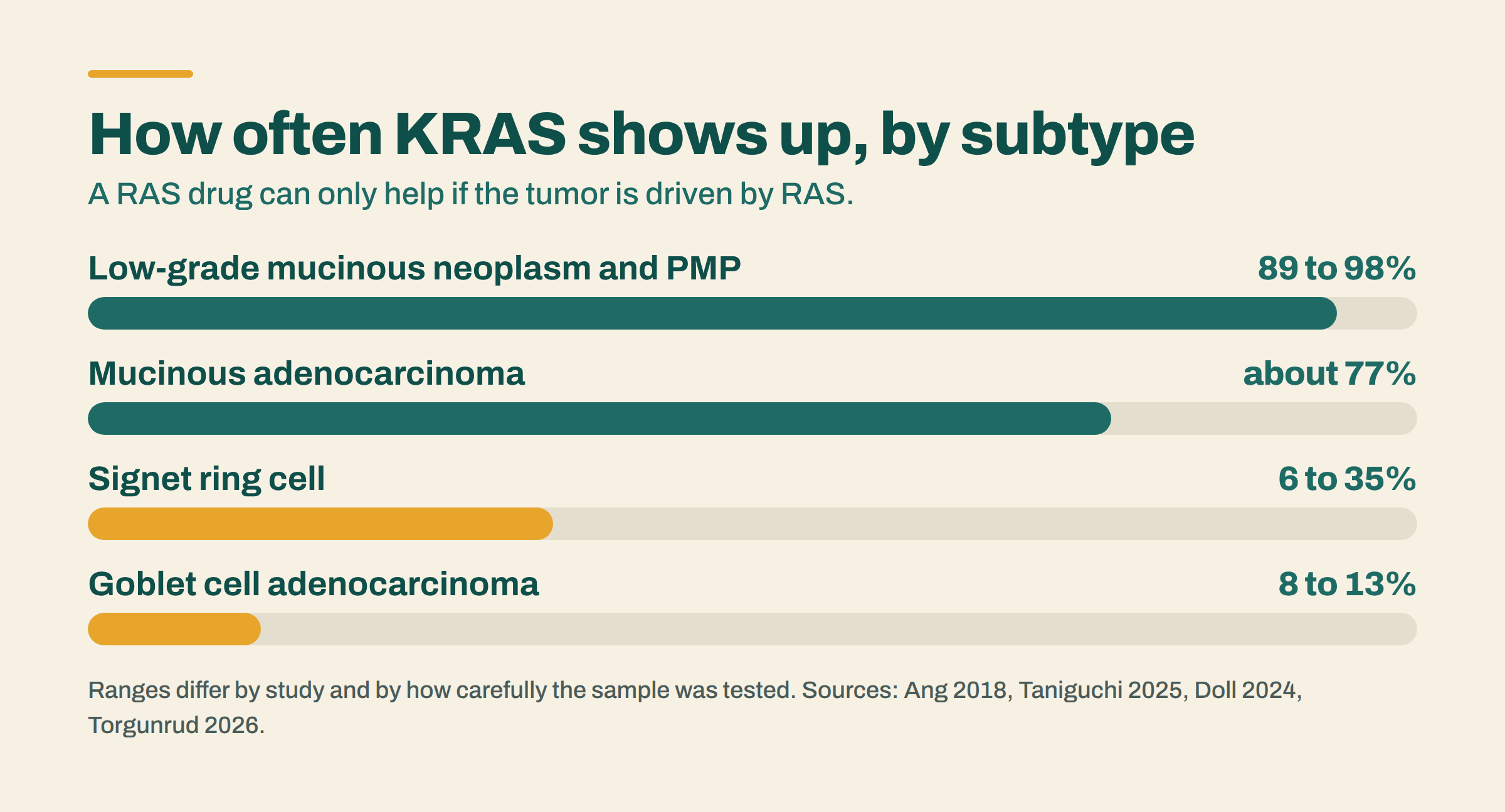
There is a detail in the Finnish study that I keep coming back to. Of the 54 tumors they found, 21 were low-grade appendiceal mucinous neoplasms and 20 were adenocarcinomas. Mucinous tumors were also the most common finding in the San Francisco study, at 54 percent.
Low-grade sounds reassuring. In this disease it is not the same as harmless. Most low-grade appendiceal mucinous neoplasms never go anywhere. But if one of them spreads or ruptures beyond the appendix and the mucin-producing cells reach the abdominal cavity, that is how pseudomyxoma peritonei can start. A lot of people in our group live with PMP.
What the research shows is narrower than a warning about what will happen. It shows that mucinous neoplasms make up a large share of the tumors found in people who first showed up with an appendix abscess. That is reason enough for our community to pay attention to this particular presentation.
Please read this part carefully
Most appendicitis is just appendicitis. Most abscesses are just abscesses. If you had an appendix abscess and it cleared up and your pathology was clean, that is good news and this post is not telling you otherwise.
What I am asking is narrower. If your appendix was never removed and examined, or if a collection near your appendix never fully went away, that is worth one conversation with your doctor. Not panic. One conversation.
Questions worth asking after an appendix abscess
- Was my appendix removed and sent to pathology, and can I get a copy of that report?
- If I was treated with antibiotics or a drain instead of surgery, was an interval appendectomy discussed, and why was it or was it not recommended?
- Did my imaging show any solid tissue inside the collection, any enlarged lymph nodes, or any nodules on the peritoneum?
- Has the collection been re-imaged since treatment, and did it actually resolve?
- Given my age and what my scan showed, is there any reason to consider a tumor here?
The honest limits
The imaging paper is two patients. It is a case report with a literature review attached, published in a new journal. The authors never state what type of cancer the first patient had. They say “appendiceal-region malignancy” and nothing more specific, so there is no way to know whether it was mucinous, goblet cell, neuroendocrine, or something else. The warning signs they list are educational. They have not been tested and validated as diagnostic criteria, and the authors say that themselves.
The tumor rate studies mostly cover mucinous neoplasms, adenocarcinoma, neuroendocrine tumors, and adenomas. They do not tell us much about goblet cell adenocarcinoma or signet ring cell disease, which show up in our community and are not well represented in these numbers. Do not stretch these percentages to cover subtypes the studies did not measure.
The Finnish study is the strongest of the group because it was prospective and ran across twelve hospitals. The San Francisco study was one center looking backward at records. Those are different levels of evidence and should not be treated as equals.
Common questions
Can an appendix abscess be cancer?
Yes. An appendix abscess can form around an underlying tumor, and the tumor can be invisible on the first scan. In a prospective Finnish study of 370 patients with an abscess around the appendix, 14.3 percent had a tumor found on pathology, compared with 1.5 percent of people with uncomplicated appendicitis.
What makes doctors suspect cancer instead of infection?
Six imaging features: a mass that does not shrink, solid tissue inside the collection that takes up contrast dye, enlarged lymph nodes, nodules on the abdominal lining, spread into unusual places, and a poor response to antibiotics or drainage.
Should the appendix always be removed after an abscess?
This is actively debated. The Finnish research team recommends that patients over 35 treated without surgery for a periappendiceal abscess go on to have an interval appendectomy. Other researchers argue that with careful selection, antibiotics alone are safe from a cancer standpoint for patients without high-risk features. Ask your surgeon what applies to your specific scan and age.
What kind of tumor is usually found?
Mucinous neoplasms are the most common, followed by adenocarcinoma and neuroendocrine tumors. Most are low grade, which is not the same as harmless, because low-grade mucinous tumors are the ones that can lead to pseudomyxoma peritonei.
Your diagnosis story is data
Nobody has counted how many appendix cancer patients were first told they had an infection or an appendix abscess. The Patient-Led Global Appendix Cancer Registry is how we start counting. It takes about twenty minutes, it is IRB approved and exempt, and it belongs to patients.
Read more on Appendicure
Why Appendix Cancer Is So Often Missed, and What Could Change That
Appendix Cancer for Emergency Physicians
Your Voice Can Help Change How the World Diagnoses Appendicitis
Sources
- Montatore M, Masino F, Tupputi R, Muscogiuri E, Guglielmi G. Cancer-Abscess: Multiple Imaging for the Early Identification of Underlying Malignancies and Differential Diagnosis with Presumed Infectious Collections. J Interdiscip Res Appl Med. 2026;6:13. doi:10.3390/jdream6030013
- Salminen R, Alajääski J, Rautio T, et al. Appendiceal Tumor Prevalence in Patients With Periappendicular Abscess. JAMA Surg. 2025;160(5):526-534. doi:10.1001/jamasurg.2025.0312
- Foroutani L, Wang JJ, Kiran S, et al. True Incidence of Appendiceal Neoplasms in Patients Presenting with Complicated Appendicitis: A Call for a Revised Management Algorithm. Ann Surg Oncol. 2026;33(5):4161-4168. doi:10.1245/s10434-025-19032-8
- Germes M, Collard MK, Laroche S, et al. Oncologic Risk of Missed Appendiceal Tumors in Acute Appendicitis. JAMA Surg. 2026;161(5):528-535. doi:10.1001/jamasurg.2026.0510
- Laudon AD, Beaulieu-Jones BR, Duraiswamy S, et al. Incidental appendiceal neoplasms in a multicenter registry of appendicitis management. Surgery. 2026;190:109879. Published online November 18, 2025. doi:10.1016/j.surg.2025.109879
- Kolbeinsson HM, Choudry HA. Editorial: True Incidence of Appendiceal Neoplasms in Patients Presenting with Complicated Appendicitis. Ann Surg Oncol. 2026. doi:10.1245/s10434-026-20487-6
- Collard MK, Challine A, Voron T. Letter to the Editor. Ann Surg Oncol. Published July 20, 2026. doi:10.1245/s10434-026-20259-2 Reply by Foroutani L, Li TM, Adam MA. Published August 3, 2026. doi:10.1245/s10434-026-20263-6
Study records located through PubMed.
This post is for education. It is not medical advice and it is not a substitute for your care team.
Appendicure runs on donations from people who understand why this matters. You can support the work here.